
Myasthenia gravis (MG) is a neuromuscular disease associated with abnormal neuromuscular (NM) transmissions.
During the sequences that take place in normal NM transmission, acetylcholine is a chemical messenger that plays a huge role in the NM junction, joining up with the acetylcholine receptors (AChRs), leading to action potential in the muscle fibre plasma membrane; therefore, movement of the muscle.
Abnormal transmissions between the nerve and muscle can lead to reduced muscle contraction that leads to clinical signs such as muscle weakness (Panel 1), which can have life-threatening effects, such as megaoesophagus and aspiration pneumonia (AP).
Panel 1. Clinical signs of myasthenia gravis
- Weakness
- Stiff gait
- Muscle tremors
- Exercise-induced weakness
- Regurgitation of food and water
- Excessive drooling
- Laboured breathing
- Dysphonia
- Decreased palpebral reflex
- Dysphagia
Two types of MG exist:
- congenital
- acquired
Congenital
Congenital MG is defined as an inherited disorder and is the rarer form of MG (Garosi, 2013).
Patients are born with fewer AChRs, meaning NM transmissions are compromised. Dogs can be as young as 6 to 12 weeks old when presented with clinical signs such as weakness (Garosi, 2013). Unfortunately, patients with congenital MG have a poor prognosis.
Acquired
Acquired MG is the more common form of MG that has been reported in breeds such as akitas, terriers, shorthair pointers and Chihuahuas (Garosi, 2013).
This form is due to an abnormal immune system, which produces antibodies to the AChRs that attack and destroy AChRs. The reason for the destruction is unknown, but results in failure of NM transmissions.
The typical presentation of a patient with acquired MG will show muscle weakness during exercise, but improving after rest (Garosi, 2013).
Diagnosis
It is important a correct diagnosis is established in MG patients, as clinical signs can often mimic myopathy and neuropathy.
Basic diagnostic testing includes thoracic radiographs to check for signs of AP and megaoesophagus, a complete blood count, and a biochemistry profile including electrolytes and urinalysis to rule out other potential causes of weakness, such as hypoglycaemia (Garosi, 2013).
Patients with suspected congenital MG would be diagnosed by having electrodiagnostics; specifically, repetitive nerve stimulation, which would show if the patient responds when the nerve is stimulated. Another diagnostic test would be to take a muscle biopsy of the external intercostal muscle (Garosi, 2013).
Acquired MG diagnosis can be achieved by a diagnostic challenge test with edrophonium chloride. The drug binds to acetylcholinesterase temporarily, which slows down the ability to hydrolyse acetylcholine, allowing more acetylcholine to remain in the synaptic cleft longer.
Ultimately, if clinical signs of weakness resolve in 15 to 20 minutes after administration of the drug, this suggests a diagnosis of MG (Merrill, 2012).
Serologic testing is a gold standard diagnostic test for acquired MG patients for circulating antibodies to the AChR. Patients on steroids must not be taking them two weeks prior to testing as results will be invalid (Merrill, 2012).
Nursing care
VNs play a pivotal role in nursing MG patients, which is challenging, but rewarding. Understanding the pathophysiology of the disease is important in helping to facilitate the best possible outcomes for the patients.
MG patients can acquire complications such as AP and megaoesophagus. It is essential veterinary nurses know how to identify these complications and how they can be prevented through the correct nursing care.
Patient monitoring
Patient monitoring it vital for MG patients. Parameters to monitor should include mentation, respiration rate and effort, membrane mucous (MM) colour, heart rate and temperature.
AP can occur if the patient inhales regurgitated stomach contents, and inflammation is caused in the alveoli due to the aspirated contents leading bacteria to potentially colonise within the lungs.
Clinical signs of AP include coughing, dyspnoea, tachypnoea, harsh lung sounds and cyanotic MM colour (Rosewell, 2015); therefore, it is imperative to be auscultating the lungs, checking MM colour, and monitoring respiration rate and effort. Patients with AP should be treated with IV antibiotics, oxygen supplementation, nebulisation and coupage.
Nutrition and hydration
Patients with MG will require supportive feeding and drinking from the VN. Food should be rolled into small bite-size balls; this can be using a wet-based food.
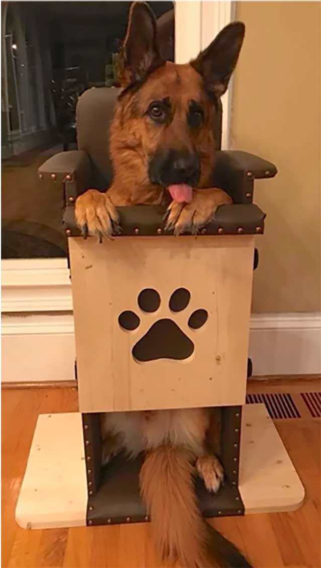
Feeding from a height is crucial in MG patients due to the risk of AP if megaoesophagus is suggested. This can be achieved by placing the patient into an upright position for 5 to 10 minutes (Figure 1) or an upright chair (Figure 2) can be used to do this. Water should also be offered from a height or on a stand to prevent aspiration.
Any signs of regurgitation should be identified with prompt notification to the vet.
IV fluid therapy may be administered under the direction of the case vet at maintenance rate (2ml/kg/hr) to maintain hydration status while hospitalised.
Oxygen therapy
MG patients may benefit from oxygen therapy. This should be done in an oxygen kennel big enough to prevent stress, and temperature should be monitored in the kennel to ensure the patient does not become hyperthermic.
Flow by oxygen can be administrated if the patient can tolerate this method. A pulse oximeter is a non-invasive method in ensuring the patient has sufficient oxygen supplementation.
Humidification
A humidifier can be used to pass oxygen through the reduction of water into steam. Humidification can be performed prior to percussion to help aid the break-up of secretions.
Patients can be nebulised for approximately 10 minutes if tolerated.
Physiotherapy
Respiratory physiotherapy can aid patients with AP and be a combination of nebulisation, percussion and vibration. Once nebulisation has been achieved, percussion in the form of coupage can be carried out for approximately 10 minutes, three to four times per day.
Secretions can become loose when vibration is expressed. The physiotherapist will create a shaking action over the chest wall during the patient’s expiration (Rosewell, 2015).
Patients with MG suffer with muscle weakness, which may result in recumbency.
The patient’s kennel should include an orthopaedic mattress for comfort, and the veterinary nurse should check the patient around its bony prominences for decubital ulcers and pressure sores.
Patients struggling to stand should be supported. This can be achieved using a sling and the patients should be taken outside for mental stimulation, urination and defecation purposes, and to prevent muscle atrophy and joint stiffness.
Conclusion
MG is a condition that has a poor prognosis due to the secondary complications that can occur, such as AP and megaoesophagus. VN care for these patients is essential, and could prevent complications and further deterioration.
VNs should be trained for physiotherapy techniques, which could aid the expulsion of secretions and any aspirated contents caused by AP.
Diagnostic testing plays a huge role in definite diagnosis and can be used in beginning treatment promptly.
Additionally, feeding remains an essential element in ensuring patients do not aspirate causing further risks to patient health, in which VNs play a vital role preventing complications.
Leave a Reply