
Skin disease is a very complex condition as many factors must be considered and evaluated to identify the cause. These will include a detailed history, thorough physical examination and appropriate diagnostic tests.
The most common causes of skin disease in dogs and cats are:
- atopic dermatitis
- allergic dermatitis, which can be broken down into food, environmental and contact
- flea allergy dermatitis
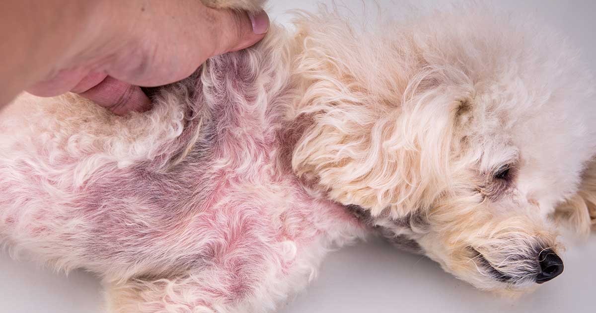
- Malassezia
- parasitic and fungal disease
Atopic dermatitis
Atopic dermatitis (AD) is defined as “a common pruritic (itchy) skin disease characterised by disturbances in epidermal (skin) barrier function that can become chronic”1. It affects both dogs and cats “with a reported frequency of 10% to 15% in dogs and around 12.5% in cats”1.
“AD is considered a hypersensitivity disorder in pets because it is associated with an exaggerated production of IgE in response to environmental allergens”1.
AD is a multifactorial disease with a genetic predisposition. Breeds predisposed to AD are “West Highland white terriers, Labrador retrievers, golden retrievers, Staffordshire bull terriers, Shar Peis and English setters, among others”2.
With cats the breed predisposition is a smaller selection. “A predisposition for Devon rex, Abyssinian and domestic shorthaired cats was reported in another study”3.
The typical clinical signs seen in dogs start to develop “between six months and three years of age”3. In cats, though, “the disease onset can vary, but commonly it is under three years”3.
The common clinical signs in dogs are:
- erythema
- pruritus
- excoriation
- pustules
- papules
- alopecia from self-trauma
- crusts
- lichenification
- saliva staining
- paronychia
- chin acne
- otitis externa
These are typically seen in the following areas: “axillae, ventral abdomen, distal extremities, inner pinnae and periocular, perioral and perianal regions are commonly affected. Otitis externa is present in half of the dogs with AD”3.
The most common clinical signs of skin disease in cats are:
- facial pruritus
- excoriation
- self-inflicted alopecia
- miliary dermatitis
- eosinophilic plaque
- eosinophilic granuloma
Cats present differently to dogs, so this should be taken into account when trying to determine the diagnosis.
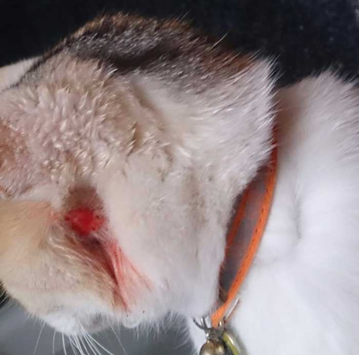
“Clinical manifestation of allergy in cats are not as site-specific as in dogs; for example, a cat scratching the neck may have a flea allergy, as well as food allergy, or a cat licking its belly may have a flea allergy, food allergy or atopic dermatitis”4. Figure 1 shows excoriation to a cat’s face.
Research has shown “the connection between AD and the gut microbiome is now being considered as a potential tool to treat the disease. After all, AD is an allergic skin disease caused by a dysfunctional immune system, and 70% to 80% of immune cells reside in the gut”1.
Allergy
It is important to try to identify the allergen. It may be helpful for a client to keep a diary of when the animal has clinical signs and in what order to identify what has changed. For example, washing powder, carpet vacuum powder, date flea treatment was applied and what brand, diet brand the animal is on, new shampoo, when the pet was last bathed and cleaning products, such as carpet shampoo and floor cleaner, as well as new plants, whether the pollen count is high and so on.
It is useful to get clients to fill in a questionnaire, and to map lesions to help chart the progress and identify causes.
Allergy testing can either be done by taking a serum blood sample or an intradermal test (the latter is usually done by a specialist).
Allergy testing is an essential tool used to help identify the causes, but it is also essential to explain to the client the importance of flea treatment and minimising exposure to potential allergens, such as house dust mites.
The patient may be able to cope with one allergen on its own, but when they start to add up – for example, AD, food allergy and flea allergy – it can become unbearable for the patient. So, we need to minimise the allergens to a level that they can cope with.
Treatment depends on the cause, so symptomatic treatment – for example, atopy and skin infection/inflammation treatment, followed by investigation to find the underlying cause. New treatments exist that block the itch receptors, or injections are available that are monoclonal antibodies, which mops up the interleukin targeting the itch at its source. This is because it is immunotherapy converting the IgE to IgG vaccine response.
Allergies can be broken down into three categories:
- food
- environmental
- contact
As previously mentioned, it can be difficult to identify the cause, so a diary is helpful to keep, followed by a map lesion. To help identify whether it is a food allergy, an eight-week food elimination diet should be performed. Ideally, you should use a source of protein and carbohydrate that the animal has not come into contact with before, such as venison and pea, or a hydrolysed diet that ensures the protein source is unrecognisable to the animal’s body. The patient must also have no other food sources, including treats, toothpaste and so on.
The patients that improve during the elimination diet then need to go on to be “challenged with individual ingredients suspected of eliciting the adverse reaction for one to three weeks”5. If the patient starts displaying symptoms again then a food allergy has been identified.
It is more difficult to conduct an elimination trial for environmental allergies. The house can be treated for house dust mites by using a house insecticide spray, airing the rooms and damp dusting.
Storage mites can be eliminated by proper storage and handling of dried food. The first step is to start a routine flea treatment programme for all the animals in the household and to treat the house for fleas.
As previously mentioned, a questionnaire, diary and map lesion should be recorded. Blood tests can be used to identify whether allergens are internal, such as dust mites, or external, such as pollens.
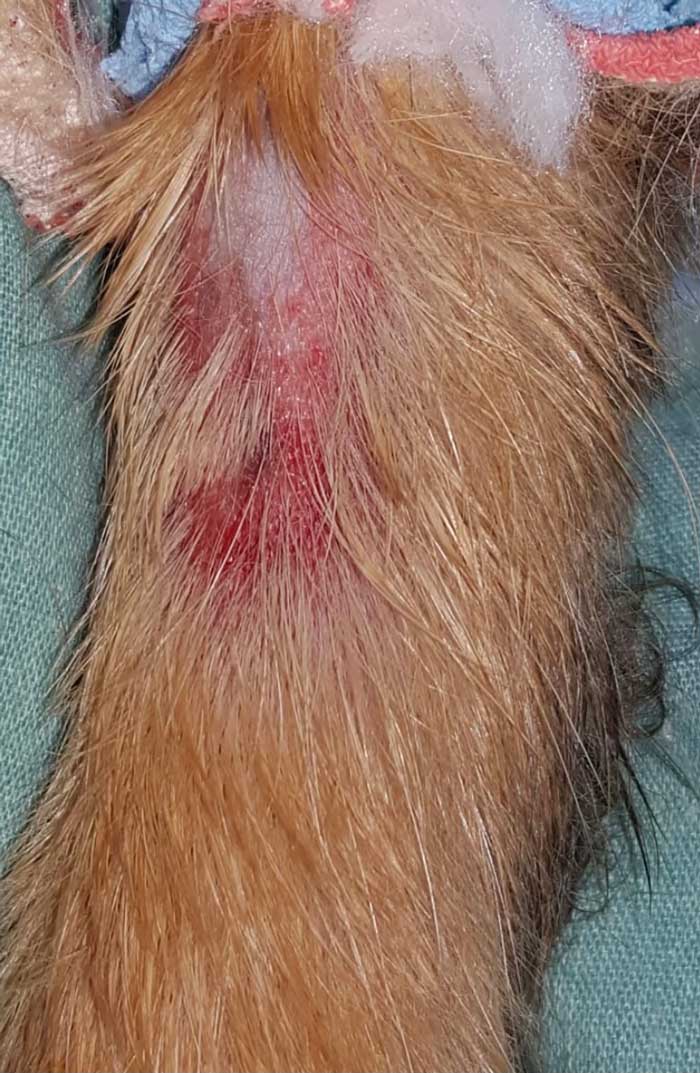
Contact allergies are something the animal has come into direct contact with. They can be “allergenic or irritant in origin, which cannot be distinguished in clinical cases”5. As previously mentioned, this could be due to anything, so a questionnaire, diary and map lesion should be recorded. A good example of this in practice is a reaction to fabric plasters (Figure 2).
Flea allergy dermatitis is an allergic reaction to the flea’s saliva. It affects both dogs and cats. “Flea allergy is not age-specific and might occur as early as three months of age”5.
Flea allergy dermatitis is usually diagnosed based on the clinical signs, and the presence of fleas and flea excrement, obtained with the wet piece of tissue and flea comb method. It generally affects “the caudal part of the body (lumbosacral area, base of tail, caudomedial thighs and abdomen) in both cats and dogs”5.
Parasites
It is important to rule out parasitic skin disease, as treatment is often curative and failure to treat will undermine the other treatments of the dermatitis.
Parasites can cause skin disease; recommended treatment will depend on the parasite found. It is quick and relatively easy to acquire a sample and diagnose it in house; for example, hair pluck, skin scrape and sticky tape impression.
These are the most common parasites found in cats and dogs:
- Sarcoptes scabiei (zoonotic)
- Demodex canis
- Notoedres
- Otodectes
- Cheyletiella blakei
- Cheyletiella yasguri
- Cheyletiella parasitovorax (zoonotic)
- Trombicula autumnalis
- fleas
- ticks (zoonotic)
- Linognathus setosus
- Trichodectes canis
Treatment will depend on the diagnosis and appropriate treatment for that species of animals.
Malassezia
Malassezia is a type of yeast found on the surface layer of the skin in healthy dogs and cats. It usually causes no problems. It is generally found “on the skin, in the ear canals and on the mucosal surfaces (oral, anal, vaginal)”6.
The most common species found is Malassezia pachydermatis. “Predisposing factors for Malassezia proliferation and pathogenicity include increased environmental humidity and temperature, skin trauma, sebum quantity and quality, immune dysfunction and genetic predispositions”7.
The breeds that are predisposed are basset hounds, American cocker spaniels, boxers and West Highland white terriers, and in cats, the Devon rex and sphinx.
Malassezia presents as itchy, scaly, inflamed skin usually seen around the lips, ear canals, neck, armpits, and between the toes and skin folds.
Patients with a severe Malassezia have a distinct smell of yeast. When chronic skin inflammation has been going on for some time, the skin becomes thickened and the skin can become darker due to excessive pigmentation. “Malassezia infection can also result in a reddish-brown discolouration of the claws”6.
Malassezia has a distinctive identifying feature – the characteristic footprint identified on microscopic examination. The sample can be taken by tape impression or swabs depending on the area, as it can be difficult to do an impression smear in some areas.
Antifungal medication is required to lower the yeast to a normal level – “it is also vital to address any underlying triggers for why the yeast numbers increased in the first place”6.
For most cases of Malassezia dermatitis, antifungal topical shampoos are effective. Alternatively, antifungal wipes, creams and fungicidal ear drops can be used for ear infestations.
In some circumstances, use of oral medication is required to treat Malassezia. “These drugs are not licensed in the UK for the treatment of Malassezia in dogs or cats, so should be reserved for cases not responding to other treatments”6.
The presence of small quantities of Malassezia is normal in cats and dogs; it is when there is an underlying condition that overgrowth can occur. “Recurrence of the problem is very common unless the underlying trigger has been identified and corrected”6.
Fungal disease
Dermatophytosis (ringworm) is a common fungal skin condition; it is zoonotic and it affects many species. “Disease is more common in young or stressed individuals, such as those in extremely crowded environments”8.
Clinical signs are hair loss, scaling, crusting, erythema, papules, hyperpigmentation and pruritus.
Dermatophytosis can mimic any pattern of skin lesion, so it should always be on the list of differential diagnoses. Care should be taken due to the zoonotic risk and appropriate PPE should be worn.
Diagnosis can be performed by examining the hair or skin by several methods, such as coat brush with a sterile toothbrush, hair pluck performed with sterile instruments such as forceps, skin biopsy, skin scrape, dermoscopy, Wood’s lamp and PCR testing.
“Skin biopsy is not indicated in routine cases of dermatophytosis. It is indicated when there are nodular lesions or an unusual presentation of a skin disease”8.
A fungal culture will determine whether spores are present on the coat. “PCR testing confirms the presence or absence of fungal DNA on the hair coat. It cannot distinguish between viable and non-viable spores”8.
It is vital to ensure all equipment used is sterile to prevent any cross-contamination and, as previously mentioned, correct PPE should be worn due to the zoonotic risk. It is also important to ensure owners are aware of the zoonotic risk and wear appropriate PPE as well.
Treatment consists of topical antifungal treatments; oral antifungal treatment, as previously mentioned, are off licence. “Topical therapy is required because it disinfects the hair coat. This is important because infective spores are the source of contagion and transmission, and disinfection minimises environmental contamination”8.
RVN’s role
RVNs can play a major role in assisting the work-ups of skin cases. RVNs can run dermatology clinics to assist the vet with case work-up following the initial consultation with the vet. This helps greatly, as it can be very time consuming for the vet, and it uses the nurse’s skill following Schedule 3 procedures.
Procedures that can be done during nurse-led dermatology clinics are: coat brushing with a toothbrush for culture, skin history questionnaire, map lesion, sticky tape impressions, comb for fleas and flea dirt, photography of lesions, hair pluck, swabs, biopsy, examination of hair (for example, broken hair from self-trauma), Wood’s lamp, skin scrapes and in-house identification under microscope.
As previously mentioned, all procedures should be performed using sterile equipment and appropriate PPE to minimise cross-contamination and exposure to zoonotic diseases.
RVNs should be used to their full potential, to help offer the patients the best service possible.
Leave a Reply