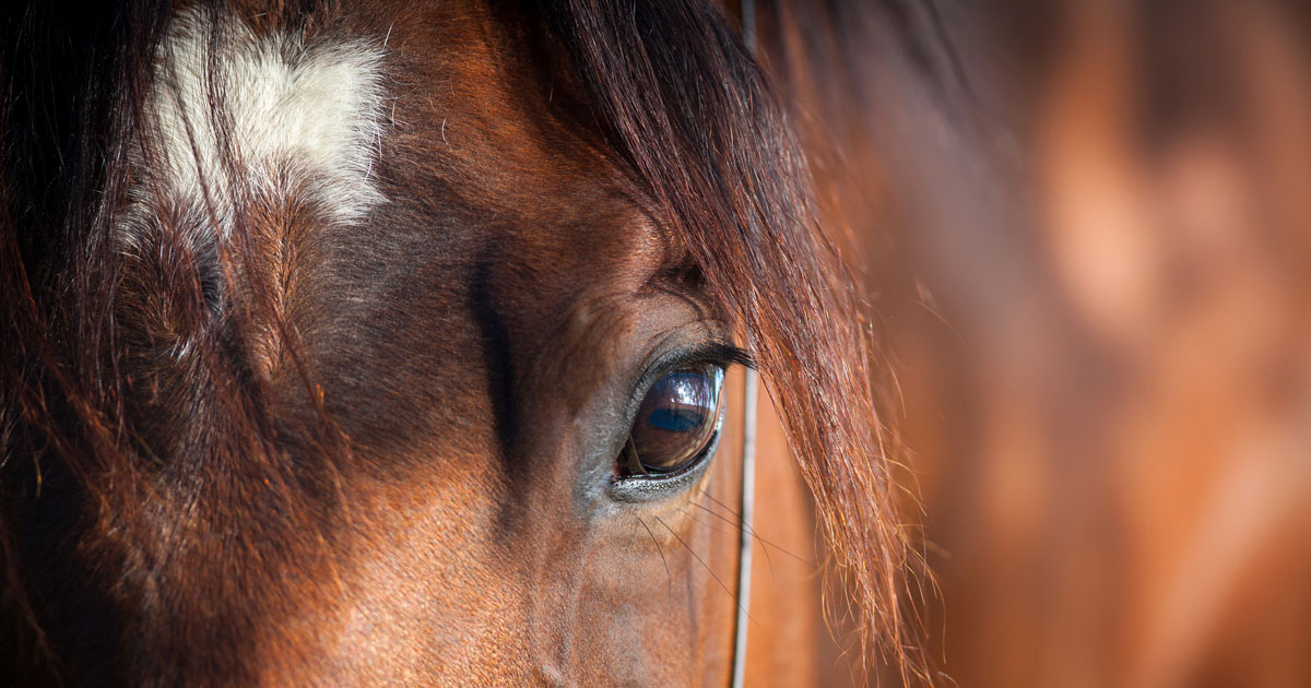
This article summarises the options available for analgesia and anaesthesia in horse. Due to surgical advances, more types of surgical procedures are being performed, both in standing horses and under general anaesthesia. Examples of anaesthetic and standing sedation protocols, as well as analgesic protocols, are presented in tables for ease of reference.
The pathophysiology of pain is discussed alongside the mechanism of action of various types of analgesic drugs and modalities used in horses. Alternative routes, such as intra-articular or epidural administration, is presented. The available analgesic drug classes include NSAIDs, spasmolytics, α2-adrenergic agonists, opioids, local anaesthetics, calcium channel α2δ-ligands, N-methyl-D-aspartate receptor antagonists, bisphosphonates, corticosteroids and the electrolyte magnesium. Various analgesic modalities such as interleukin-1 receptor antagonist proteins (IRAP), acupuncture and electro-acupuncture, mesotherapy and neurectomy are also discussed.
Choosing the right analgesic or anaesthetic drug, dose, route, frequency or duration relies on our understanding of physiological and pathophysiological mechanisms at play in each patient.
Individual patient factors (for example, temperament or underlying disease), potential side effects, cost, evidence of efficacy, and regulatory considerations also play an important role. Due to the limited number of drugs licensed for use in horses, equine practitioners need to consider additional compounds under the prescribing cascade to improve the quality and safety of anaesthesia and analgesia.
The majority of the drugs mentioned in this article are not licensed for the purposes discussed, but literature references are provided1,2.
Anaesthesia and standing sedation
While the available anaesthetic and analgesic drugs have not changed in decades, the way we use them has changed significantly.
To prevent risks associated with general anaesthesia, and due to advances in equine surgery, many surgical procedures are now performed in standing horses, necessitating ever-improving standing sedation protocols.
Using a balanced approach that relies on diverse mechanisms improves the safety and efficacy of anaesthetic and analgesic protocols.
According to an international survey, acepromazine, xylazine, detomidine, butorphanol, ketamine, diazepam, isoflurane, lidocaine, phenylbutazone and flunixin meglumine are the drugs most commonly used in equine general anaesthesia3.
Equine anaesthesia is divided into premedication, induction, maintenance and recovery. Premedication consists of sedation with an α2-adrenergic agonist, as well as additional analgesia, antibiotics and anti-inflammatories, if warranted by the procedure. Induction is most commonly achieved with a combination of ketamine and a muscle relaxant, such as midazolam or guaifenesin.
Maintenance of anaesthesia can be achieved with volatile anaesthetic agents, such as isoflurane, halothane, sevoflurane and desflurane, but all result in significant cardiorespiratory suppression and fail to provide analgesia. The use of partial IV anaesthesia (known as PIVA) and total IV anaesthesia (TIVA) decreases cardiopulmonary suppression associated with inhalant anaesthetic drugs and provides analgesia.
Continuous-rate infusion (CRI) of drugs provides improved stability of plasma drug concentrations compared to intermittent boluses, thus providing greater stability of physiological parameters.
The use of CRIs of sedative and analgesic drugs, as well as epidural anaesthesia and analgesia, facilitates protracted and painful standing procedures.
After an initial bolus, various drugs and combinations of drugs (α2-adrenergic agonists, benzodiazepines, opioids and ketamine) can be administered as a CRI, and may be combined with epidural drugs to improve analgesia and regional anaesthesia. One of the difficulties in equine anaesthesia, sedation and analgesia is the enormity of variation between patients in individual responses to drugs.
Intensive monitoring of the sedated or anaesthetised patient is, therefore, imperative to ensure adjustments in drugs and dosages are made in a timely fashion. The Ghent Sedation Algorithm that scores standing sedation based on depth, quality and ataxia was published this year4. It provides easy-to-follow recommendations for altering the rate of a CRI based on a horse’s sedation scores. Examples of drug protocols used in general anaesthesia and for standing procedures can be found in Tables 1 and 2.
| TABLE 1. Anaesthetic drugs used in horses – selected examples | |||
|---|---|---|---|
| Pre-medication | |||
| NSAID + α2-adrenergic agonist +/− opioid +/− antibiotics +/− acepromazine | |||
| Induction agents (Wohlfender et al, 2015; Lerche, 2013) | |||
| Drug | Dose | Route | Comments and references |
| Ketamine
OR Alfaxalone |
2.2mg/kg
1mg/kg |
IV | Induction agents are used together with a muscle relaxant for induction (for example, midazolam, diazepam, or glyceryl guaiacolate either) after pre-medication with an α2-adrenergic agonist +/− opioid.
(Kloppel and Leece, 2011) |
| Diazepam
OR Midazolam OR Guaifenesin |
0.04mg/kg to 0.1mg/kg
0.04mg/kg to 0.1mg/kg
35mg/kg to 50mg/kg |
IV
IV
IV infusion |
All three drugs provide muscle relaxation during anaesthetic induction and decrease inhalant anaesthetic minimum alveolar concentration (MAC). Diazepam or midazolam can be mixed with ketamine in a syringe. Ketamine administered as soon as the horse becomes ataxic from guaifenesin (knees start to buckle). |
| Drugs used to decrease inhalant anaesthetic MAC in partial IV anaesthesia protocols (Gozalo-Marcilla et al, 2014, 2015; Valverde, 2013) | |||
| Protocols | Comments and references | ||
| Lidocaine 1.3mg/kg loading dose over 15 min, then 50μg/kg/min CRI. | Stop 20 to 30 min prior to recovery to avoid ataxia. Decreases sevoflurane MAC by 30%. | ||
| Ketamine 0.5mg/kg/hr to 3mg/kg/hr CRI (1mg/kg/hr recommended). | Approximately 20% reduction in isoflurane MAC with 1mg/kg/hr CRI. | ||
| Xylazine 0.6mg/kg loading dose, then 1mg/kg/hr CRI. | Approxomately 20% reduction in isoflurane MAC. | ||
| Medetomidine 3.5μg/kg to 7μg/kg loading dose, then 1.75μg/kg/hr to 5μg/kg/hr. | Has a short half-life (ideal for CRI), improves blood pressure (Sacks et al, 2017; Kempchen et al, 2012). | ||
| Medetomidine 3.5μg/kg to 7μg/kg loading dose, then 1.75μg/kg/hr to 5μg/kg/hr. | Has a short half-life (ideal for CRI), improves blood pressure (Sacks et al, 2017; Kempchen et al, 2012). | ||
| Lidocaine 2mg/kg loading dose, then 3mg/kg/hr PLUS Ketamine 3mg/kg/hr CRI PLUS/MINUS Morphine 0.15mg/kg bolus, then 0.1mg/kg/hr | Decreases isoflurane MAC by 49% to 53% (Villalba et al, 2011). | ||
| Selected examples of TIVA protocols (Valverde, 2013) | |||
| Premedication with α2-adrenergic agonist + induction with ketamine 2.2mg/kg +/− midazolam 0.05mg/kg to 0.1mg/kg. | 15 min surgical anaesthesia, recovery typically after 20 min to 25 min. Surgical time can be extended by “topping up” with 30% to 50% of the α2-adrenergic agonist and ketamine dose at the first movement. | ||
| “Triple drip”: 1,000mg to 2,000mg of ketamine + 500mg of xylazine in 1L of 5% guaifenesin and administered to effect up to 2ml/kg/hr. | Used to maintain field anaesthesia (shorter procedures) after premedication and induction. Not suitable for procedures lasting >1 hour. | ||
| 1,000mg ketamine + 500mg xylazine + 50mg midazolam are added to 1L of isotonic fluids and administered at a rate of 2.2ml/kg/hr. | Used to maintain anaesthesia after premedication and induction. If anaesthetic duration is >45 min, then oxygen supplementation and ventilation are imperative. | ||
| Alfaxalone2mg/kg/hr to 3mg/kg/hr. When necessary, additional alfaxalone boluses (0.2mg/kg) can be administered IV. | Used to maintain anaesthesia after premedication and induction (Deutsch et al, 2017; Goodwin et al, 2019). | ||
| Table references are listed in “Further Reading” below | |||
Analgesia: the pain problem
Mechanical, thermal, chemical or electrical noxious stimuli induce transient receptor potentials in multimodal high-threshold nociceptors. Action potentials are then transmitted to the spinal cord along nerve fibres. Myelinated rapidly transmitting A-δ fibres carry information about noxious deforming mechanical stimulation.
C-polymodal fibres are unmyelinated, slowly conducting nerve fibres that carry information about mechanical, thermal and chemical noxious stimuli.
Noxious chemicals include endogenous substances such as inflammatory mediators, or exogenous chemicals (for example, acids, caustics). These fibres synapse in the spinal cord on dorsal horn neurons, and pain is transmitted along the spinal cord to the brainstem and brain through the dorsal column neurons and the spinothalamic tract5,6.
Visceral pain is conducted exclusively by C fibres of the autonomic nervous system, which is why it is associated with relatively greater physiologic responses, such as tachycardia, hyperpnoea, hypertension and sweating7. To achieve analgesia, the pain pathway needs to be interrupted or modulated, which can be done at multiple levels.
Tissue damage causes sensitisation of peripheral nociceptors, which leads to allodynia and hyperalgesia. This change in nervous system function is mediated through the release of many substances, including pro-inflammatory cytokines (interleukin-1, interleukin-6 and interleukin-8, and tumour necrosis factor alpha [TNFα]), products of the arachidonic acid cascade (prostaglandins and leukotrienes), the sympathetic nervous system (noradrenaline), substance P, calcitonin gene-related peptide, nitrous oxide, nerve growth factors, histamine, H+ and K+6,8.
Nociceptors become sensitised to noxious and even non-noxious stimuli through a decrease in activation threshold, which leads to hyperalgesia. Inactive nociceptors are also recruited to transmit pain signals causing allodynia9.
Persistent or extreme pain can cause adaptive or maladaptive changes in the sensory nervous system, leading sensitised central and peripheral nociceptors to release pain mediators in the spinal cord (for example, prostaglandins, substance P and glutamate), which activates NK-1 and N-methyl-D-aspartate (NMDA)/α-amino-3-hydroxy-5-methyl-4-isoxazole propionic acid receptors and lowers the pain response threshold in the spinal cord dorsal horn neurons. This affects the way any nociceptive signal in the body is transmitted, causing widespread allodynia and central (or secondary) hyperalgesia10.
Pre-emptive use of analgesics for any procedure that can lead to pain or inflammation is, therefore, highly effective, as it prevents sensitisation, hyperalgesia and allodynia11,12.
Neuropathic pain
Excessive stimulation of nociceptive pathways or physical damage to the neurons leads to uncoupling of pain from the inciting cause. The pain therefore becomes a disease entity in its own right. Equine examples of conditions resulting in or from neuropathic pain include chronic laminitis and trigeminal neuralgia.
Laminitis pain is multifactorial and can be extremely variable between horses13. Third phalanx rotation and sinking injures the C and A-δ nerve fibres in the hoof, resulting in spontaneous impulse discharges and sustained (neuropathic) pain. This persistent peripheral nerve firing also results in central hyperalgesia9.
Neuromorphological changes and altered gene expression in horses with recurrent or refractory laminitis are strikingly similar to those that occur in animal models of peripheral nerve injury, and humans with neuropathic pain14.
Analgesics used in horses
Short-term analgesia required for surgical and standing procedures can be achieved through various combinations of oral, mucosal, IV, intrathecal, perineural or intra-articular drugs. Having a good grasp of all the different options can assist in improved restraint, clinician safety and analgesia.
When long-term analgesia is needed, we have traditionally relied on oral NSAIDs, but several additional drugs and analgesic modalities are available. Examples of drug protocols used for equine analgesia can be found in Table 2.
NSAIDs are very effective in reducing hyperalgesia due to peripheral sensitisation (for example, OA). They effectively inhibit inflammation through inhibition of the metabolism of arachidonic acid to prostaglandins, among other poorly understood mechanisms15.
The non-selective cyclo-oxygenase (COX) inhibitors phenylbutazone and flunixin meglumine remain the most commonly used NSAIDs for musculoskeletal and colic pain16. Their potential for gastrointestinal and renal side effects are well known. For the majority of horses with mild to moderate acute pain (for example, foot abscess, injuries or medical colic), NSAIDs may provide sufficient analgesia.
Choosing the optimal dose of NSAID requires some consideration. To avoid side effects, one might be tempted into trying to decrease the dose of NSAID. However, in a lameness model, decreasing the dose of flunixin from 1mg/kg to 0.5mg/kg severely limited the analgesic effect17.
Increasing the dose above recommended doses to try to provide additional analgesia is also not advisable – not only due to the increased risk of side effects. In clinical patients, increasing the dose of phenylbutazone from 4.4mg/kg to 8.8mg/kg did not provide additional analgesia18.
Co-administration of NSAIDs (2.2mg/kg phenylbutazone PO plus 1.1mg/kg flunixin PO every 12 hours) improves clinical lameness beyond the effect of phenylbutazone alone19. Co-administration of the non-selective drugs with paracetamol can also be considered. The more COX-2 selective drugs carprofen, meloxicam and deracoxib, or the COX-2-specific drug firocoxib, may have fewer side effects20-23, but their availability and cost can make chronic use impractical.
As with all analgesics, sedatives and anaesthetics, individual horses may have vastly different drug responses.
If a horse has an unexpectedly poor response to one NSAID, it may be worth trying a different compound. This variability is likely due to genetic receptor polymorphisms and is seen in many species including man.
Topical diclofenac has been shown to decrease clinical lameness24. It also decreases skin inflammation in experimental models25, and around IV regional perfusion sites26. It is capable of disease modification and improving clinical signs in horses with experimentally induced OA27, although results were not as positive in experimental synovitis28. Minimal systemic absorption in foals and adults makes the drug attractive to limit NSAID-related side effects29,30.
N-butylscopolammonium bromide is an anticholinergic and antispasmodic drug that provides analgesia in cases with non-strangulating large intestinal obstructions and spasmodic colic31-33. Administration is associated with transient tachycardia.
Dipyrone (otherwise known as metamizole) is an analgesic, anti-spasmodic and antipyretic drug suspected to inhibit central nervous prostaglandin production. It is predominantly used in combination with n-butylscopolammonium bromide, although veterinary-approved dipyrone is available on its own in certain countries.
The α2-adrenergic agonists are excellent short-term analgesics and potent sedatives. When used as CRIs, their action can be prolonged in both standing and anaesthetised patients. Side effects are well known and include peripheral vasoconstriction, bradyarrhythmia, decreased cardiac output and decreased intestinal transit34.
Due to their spinal and supraspinal effects, epidural administration is particularly effective. The duration and intensity of sedative and analgesic properties varies between drugs (see Table 2)35,36. When comparing single-drug boluses, detomidine provides the longest-lasting, most potent analgesic effect.
Romifidine produces the smallest amount of ataxia, which is beneficial to anaesthetic recovery, and is the longest acting.
Medetomidine has the shortest half-life and produces a lot of muscle relaxation (ataxia), making it ideal for use as a CRI during general anaesthesia and in combination with ketamine and/or opioids for standing restraint.
Opioids are frequently used as part of balanced sedation, analgesic and anaesthetic protocols. They have positive effects on restraint when used in combination with α2-adrenergic agonists, and potentiate the analgesic and sedative effects of these drugs, but provide inconsistently quantifiable analgesia when used on their own36,37. Side effects include variable excitation.
Opioids used in horses include pure μ-agonists (for example, morphine, methadone, fentanyl and alfentanil), partial μ-agonists (for example, buprenorphine), and κ-agonist/m-antagonists (for example, butorphanol or nalbuphine). Intra-articular, perineural and epidural administration of opioids provides effective analgesia and limits side effects.
Epidural morphine, both alone and in combination with α2-adrenergic agonists, provides effective and prolonged analgesia for painful standing and surgical procedures (for instance, laparoscopic ovariectomy or castration)38-41. Intra-articular morphine also provides lasting analgesia42,43.
Local anaesthetics are useful to facilitate lameness evaluation, standing surgery via local, regional or epidural route, and as part of balanced general anaesthesia. They exert their local and regional effects through reversible blockade of nerve fibre impulse propagation by binding to sodium channels and preventing sodium influx (depolarisation).
IV lidocaine is also anti-arrhythmic and a very effective analgesic that targets both the spinal and supraspinal nociceptive systems. Although its analgesic mechanism in horses is incompletely understood, it suppresses peripheral hyperalgesia, central sensitisation and allodynia in lab animals and humans44-46.
Additionally, lidocaine has immunomodulatory properties that appear protective against ischaemic and reperfusion injury in various species including the horse47-49.
Even when local anaesthetics are administered regionally in surgery, positive effects on anaesthesia and the systemic inflammatory response has been demonstrated in a group of horses undergoing castration.
The addition of SC, intrafunicular and intratesticular mepivacaine to a balanced general anaesthetic protocol improved anaesthetic quality, attenuated postoperative increases in cytokines and improved analgesia50.
Calcium channel α2δ-ligands, such as gabapentin, bind to the α2δ-1 subunit of voltage-gated calcium channels in the spinal cord and brain, resulting in changes to the release of neurotransmitters such as glutamate, GABA, norepinephrine and substance P in the CNS. Gabapentin is used for the treatment of neuropathic pain and as an adjunct to treatment of painful conditions with a significant hyperalgesic component, such as laminitis36, 51-54.
Among other poorly understood mechanisms, ketamine is best known as an NMDA receptor antagonist. It is most commonly used for anaesthetic induction, but is also useful in the management of severely painful conditions such as osteomyelitis, septic joints, burns and colic, as well as in combination with other drugs for standing procedures55.
The analgesic mechanisms associated with acupuncture are incompletely understood, but include local effects at needle insertion, modulation of circulating neurotransmitters and cell signalling in peripheral tissues, as well as the CNS, and effects on myofascial trigger point pathology. When performed by trained professionals, acupuncture is safe and effective, and can be considered in a multimodal pain management plan.
Although scientific study of this modality in horses is still limited, compelling evidence in basic science, and clinical research in various other species supports its safety and efficacy56-60.
The addition of electricity to acupuncture needles (electroacupuncture) has been evaluated in horses. It significantly increases skin and rectal pain thresholds in horses and also increases the concentration of β-endorphins in CSF61,62.
Mesotherapy, the intradermal injection of local anaesthetics and other substances, has long been used to treat equine back pain56. The underlying mechanism may relate to a slower spread, higher levels and longer-lasting effect of drugs in areas underlying injection sites, or could be due to disruption of pain signals in the neuro-immuno-cutaneous system (NICS).
Although a paucity of studies in horses exists, promising research is available in other species. In a well-designed human study, mesotherapy was superior to systemic therapy for pain relief of musculoskeletal injury in short-term follow-up of emergency department admissions63.
It also reduces pain and disability in humans with non-specific chronic back pain64, and was at least as good as carprofen for control of chronic back pain in 10 working dogs65. In the NICS system, signals are transmitted between neuronal and non-neuronal cutaneous cells (for example, keratinocytes) in response to tissue injury.
In neuropathic pain states, human keratinocytes have increased expression of nociceptive receptors, which increases the amount of pain perceived because these cells form tight junctions with sensory afferent fibres. This mechanism may explain why topical lidocaine patches have proven effective for pain management in humans with post-herpetic neuralgia66.
Trigeminally mediated headshaking in horses is a neuralgia caused by the reduced activation threshold of the maxillary branch of the trigeminal nerve. Both percutaneous electrical nerve stimulation and electroacupuncture have been used successfully as palliative treatments67,68. Electrical stimulation is believed to activate A-β fibres, which in turn modulate the transmission of pain signals from nociceptive A-δ and C fibres in the spinal cord69.
IV and oral magnesium, as well as oral magnesium-boron, are also promising treatments believed to decrease the trigeminal nerve activation threshold70,71.
Interleukin-1 receptor antagonist proteins are useful to decrease pain and inflammation and improve function of joints affected by OA in various species, including humans and horses56,72,73.
Combinations of systemic NSAIDs, intra-articular corticosteroids, viscosupplementation and chondroprotectants are also used in the treatment of OA74. Bisphosphonates are used to provide analgesia in OA and navicular syndrome75.
When all other analgesic and rehabilitative options have been exhausted, carefully selected cases suffering from proximal suspensory desmopathy may benefit from neurectomy and fasciotomy of the deep branch of the lateral plantar/palmar nerves76,77.
Palmar or plantar neurectomy can also improve or resolve lameness in horses with unresponsive foot pain, but advanced imaging (for example, MRI) is required to inform appropriate candidate selection78.
Conclusion
In conclusion, numerous anaesthetic and analgesic drugs and modalities are available to the equine clinician. Finding the optimal combinations of drugs, protocols and modalities for specific conditions requires further study.
| Table 2. Analgesics and anti-nociceptives used in horses – selected examples (Sanchez and Robertson, 2014) | |||||
|---|---|---|---|---|---|
| Drug | Dose | Route | Dosing interval or duration of analgesia | Most common use and selected comments | References/further reading |
| Flunixin meglumine | 0.5mg/kg to 1.1mg/kg | IV, PO | 12h to 24h | Musculoskeletal and visceral pain and inflammation, perioperative analgesia | |
| Phenylbutazone | 2.2mg/kg to 4.4mg/kg | IV, PO | 12h to 24h | Musculoskeletal pain and inflammation. Some visceral analgesia | |
| Paracetamol | 20mg/kg | PO | 12h | Adjunct to other analgesic drugs | (Mercer at al, 2019) |
| N-butylscopolammonium bromide | 0.2mg/kg to 0.3mg/kg | IV | 20 min to 60 min duration of effect | Analgesia in non-strangulating large intestinal obstruction and spasmotic colic. Rapid analgesic onset | (Roelvink et al, 1991; Boatwright et al, 1996; Sanchez et al, 2008) |
| Dipyrone/metamizole | 25mg/kg | IV | 50 min duration of effect when combined with n-butylscopolammonium bromide | Colic, chronic arthritis, spasmolytic. Can rarely cause agranulocytosis | (Roelvink et al, 1991) |
| Meloxicam 0.6mg/kg | 0.6mg/kg | IV, PO | 24h | Musculoskeletal pain and inflammation, perioperative analgesia. Rapid clearance means shorter detection times in competition horses and improved safety in foals |
(Friton et al, 2006; de Grauw et al, 2009; Naylor et al, 2013) |
| Firocoxib | 0.1mg/kg | IV, PO | 24h | Musculoskeletal pain and inflammation | (Orsini et al, 2012) |
| Xylazine | 0.2mg/kg to 1.1mg/kg | IV, IM | Bolus | Sedation and 20 min to 60 min of analgesia, premedication | |
| Detomidine | 5μg/kg to 40μg/kg | IV, IM | Bolus | Sedation and up to 90 min of analgesia, premedication | |
| Romifidine | 0.06mg/kg to 0.1mg/kg | IV | Bolus | Sedation and up to 120 min of analgesia, although less potent than detomidine, premedication | |
| Medetomidine | 4μg/kg to 10μg/kg | IV | Bolus | ||
| Detomidine
PLUS Methadone |
5μg/kg
0.2mg/kg |
IV | Bolus | Methadone potentiates the antinociception produced by detomidine with minimal sedative effects | (Gozalo-Marcilla et al, 2017) |
| Lidocaine
PLUS
Ketamine
|
1.3mg/kg
then 3mg/kh/h
0.55mg/kg then 0.5mg/kg/h |
IV | Bolus
CRI
Bolus
CRI |
Analgesia for severe pain. Can be used for several days. No benefit to adding butorphanol | |
| Paracetamol
PLUS Tramadol |
6g/h
1mg/kg then 3mg/kg/h |
IV | CRI Bolus CRI |
Provides very good analgesia. Potential for paroxysmal ventricular tachycardia | (Tavanaeimanesh et al, 2018) |
| Buprenorphine | 5μg/kg to 10μg/kg | IV | 6h to 12h | Sedation with acepromazine or α2-adrenergic agonists decreases excitation for all opioids. Buprenorphine had better postoperative analgesic scores compared to butorphanol | (Taylor et al, 2016) |
| Butorphanol | 10μg/kg to 50μg/kg
40μg/kg to 100μg/kg 18μg/kg |
IV
IM
IV
|
3h to 4h
4h to 6h
Bolus then CRI
|
Sedation with acepromazine or α2-adrenergic agonists decreases excitation for all opioids. Buprenorphine had better postoperative analgesic scores compared to butorphanol | (Taylor et al, 2016) |
| Morphine | 0.12mg/kg to 0.66mg/kg | IV | 4h to 6h | Sedation with acepromazine or α2-adrenergic agonists decreases excitation for all opioids. Buprenorphine had better postoperative analgesic scores compared to butorphanol | (Taylor et al, 2016) |
| Ketamine | 0.4mg/kg/h to 1.2mg/kg/h | CRI | Effective for severe pain. Safely shown for at least 5 days at 0.8mg/kg/h | (Wagner et al, 2011; Matthews et al, 2004 | |
| Gabapentin | 2.5mg/kg to 20mg/kg | IV, PO | 12h | For neuropathic pain or as an adjunct to treatment for severe or chronic pain leading to hyperalgesia. Poor oral bioavailability | (Dirikolu et al, 2008; Davis et al, 2007; Terry et al, 2010) |
| Morphine | 120mg | Intra-articular | Potent analgesic and variable anti-inflammatory effects | (van Loon et al, 2010; Rubio-Martinez et al, 2017) | |
| Morphine | 100mg/animal or 0.05mg/kg to 0.2mg/kg
0.1mg/kg
|
Caudal epidural
Cranial epidural
|
3h to 8h duration
>12h duration
|
Dilute to 30ml with saline. Excellent analgesia and anti-hyperalgesia. Can cause pruritis
Thoracolumbar admistration achieved via epidural catheter |
(van Loon et al, 2012; Martin-Flores et al; 2014)
(Freitas et al, 2011) |
| Lidocaine | 0.2mg/kg | Epidural | 30 min to 90 min duration | 5ml to 8ml of 2% solution. Perineal anaesthesia. Onset within 10 min. Risk of hindlimb weakness | |
| Bupivacaine | 0.04mg/kg to 0.06mg/kg | Epidural | 3.5h to 5h duration | 5ml to 8ml of 0.25% solution. Perineal anaesthesia. Risk of hindlimb weakness | |
| Xylazine | 0.03mg/kg to 0.06mg/kg | Epidural | 1h to 3.5h duration | 0.2mg/kg typical analgesia. Onset can take 30 min. Relatively less hindlimb weakness (increases with increasing dose) | |
| Detomidine | 0.02mg/kg to 0.06mg/kg | Epidural | 2h to 4h duration | Onset 10 min to 25 min analgesia | |
| Morphine PLUS Detomidine |
0.1mg/kg to 0.2mg/kg
0.01mg/kg to 0.03mg/kg |
Epidural | 24h to 48h duration | Analgesia | (Rowland et al, 2018) |
| Morphine
PLUS Xylazine |
0.01mg/kg to 0.02mg/kg | Epidural | 12h and 18h duration | Analgesia | |
| Lidocaine
PLUS Xylazine |
0.22mg/kg
|
0.17mg/kg | Epidural | 4h to 6h duration | Perineal anaesthesia for standing surgeries such as laparoscopic ovariectomy |
| Romifidine
PLUS
Butorphanol |
80ug/kg then 29ug/kg/h
18ug/kg then 25ug/kg/h
|
IV | Bolus, CRI
Bolus, CRI |
Standing restraint for surgical and other procedures (for example, dentistry and ophthalmology procedures) | (Marly et al, 2014) |
| Detomidine
PLUS Morphine |
10ug/kg then 3ug/kg/h
0.1mg/kg |
IV
  IV |
Bolus, CRI
Bolus after 5 min |
Standing surgery/procedures | (Potter et al, 2016) |
| Romifidine
PLUS
Midazolam |
30ug/kg then 50ug/kg/h
0.02mg/kg then 0.06mg/kg/h |
IV | Bolus CRIBolus CRI |
Standing surgery/procedures
|
(Muller et al, 2017) |
| Romifidine
PLUS
Ketamine PLUS |
30ug/kg then 50ug/kg/h
0.5mg/kg then 1.2mg/kg/h |
IV | Bolus
CRI
Bolus CRI |
Standing surgery/procedures |
(Muller et al, 2017) |
| Table references are listed in “Further Reading” below | |||||
Leave a Reply