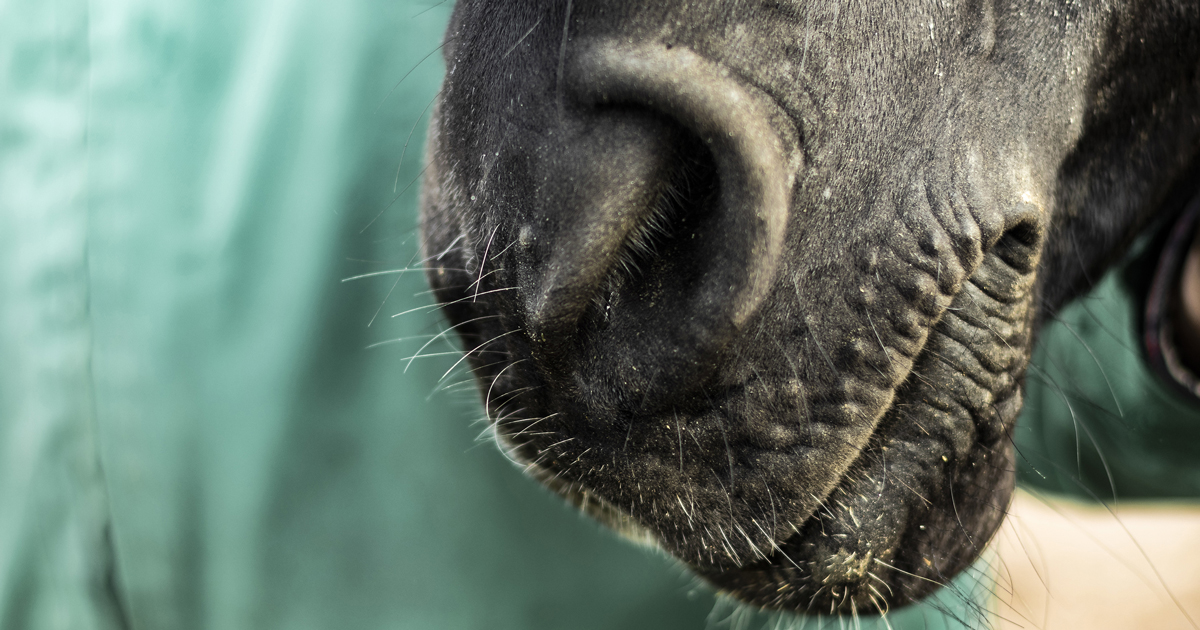
Skin disease in horses is a common problem. However, diagnosis can present a significant challenge.
RVNs do not commonly have a large role in the diagnosis of equine dermatological conditions. However, it is clear a role for RVNs does exist in this area.
This article will look at protocols associated with equine dermatology, and, where possible, how RVNs can assist with these procedures and enhance patient care.
Diagnostic techniques
Skin scrapings
Skin scrapings are straightforward and performed frequently in equine practice. However, they are often unrewarding. Samples are, by their nature, very superficial and comprise scale that will not enable diagnosis of many common equine skin conditions (Rendle, 2017).
Deeper scrapes can be performed by pinching the skin between the finger and thumb, and using a scalpel blade to scrape the skin until capillary bleeding occurs (Rendle, 2017).
Skin scrapes can be taken at the periphery of dermatophytosis (ringworm) lesions for microscopy, but with the advent of the dermatophyte PCR diagnosis (which is more sensitive and specific), this technique has been superseded (Prutton, 2019). RVNs can assist with this procedure by taking samples themselves and maintaining infection control protocols. This is particularly important with dermatophytosis as it is a zoonotic disease.
Using the correct personal protective equipment (PPE) and correctly disinfecting any equipment is paramount to prevent the spread of disease to other patients and human colleagues.
Acetate tape impressions
Performing tape impressions is quicker and more straightforward than skin scrapings. Acetate tape is applied with gentle pressure to collect material from the skin surface. The tape is then applied gently to a microscope slide without pressure and viewed under the microscope.
Tape impressions are particularly useful and would be the test of choice for the identification of Oxyuris equi (pinworm) eggs from around the anus (Rendle, 2017).
Tape impressions can also be performed in some cases of Chorioptes mites and Damalinia equi lice infestations, but a negative result does not rule out an infection. If ectoparasites are ruled out, or considered clinically unlikely, further diagnostics can be undertaken and will include culture, PCR, biopsy and intradermal skin tests (Prutton, 2019).
RVNs can take samples using acetate tape and evaluate them under the microscope to identify parasites if they are present. The results would then be reported back to the case vet. Once a diagnosis has been made, the RVN can assist by administering treatment – for example, bathing the patient with medicated shampoo, or administering topical or IV medications as prescribed by the case vet. The patient would also need to be monitored for progress and any observations would be reported back to the vet.
Hair plucks
Hair plucks can be useful in the diagnosis of dermatophytosis with the advent of PCR, and should be taken from the periphery of the lesion – with multiple (greater than 20 to 30) hairs taken and stored in a container (Prutton, 2019).
Due to the infectious nature of dermatophytosis, infection control is very important. RVNs can help to ensure the hairs are stored in an appropriate container to reduce exposure to laboratory staff.
All equipment involved will need to be disinfected correctly. This is something an RVN can get involved with, as well as advising the owner of the patient on correct disinfection of fomites, and the stable environment at home.
Bacterial and fungal culture
Where bacterial infection is suspected, it is appropriate that swabs or tissue samples are collected and submitted for bacterial culture (Rendle, 2017). If swabs can be collected from pustules or discharging tracts without contamination from the skin surface, then this is appropriate.
Aseptic preparation of the skin surface prior to sampling deep lesions will reduce the risk of confounding results with contaminating or commensal bacteria from the skin surface (Rendle, 2017).
The RVN can assist with the sterile preparation of the site. First, the area will be clipped carefully and neatly. Wearing sterile gloves, the RVN can then scrub the area using 4% chlorhexidine, making sure there is at least a five-minute contact time. The area should then be rinsed with sterile saline ready for the sample to be taken.
If pustules or lesions consistent with folliculitis are present, then punch biopsies can be collected following aseptic preparation of the skin surface and macerated into a suitable media prior to bacterial culture (Rendle, 2017).
The RVN can again assist with the sterile preparation of the area, prepare the equipment required, and make sure a suitable sample pot is available. Bacterial culture of superficial skin scrapes is rarely rewarding and more likely to yield growth of contaminants or commensals of no clinical relevance (Rendle, 2017).
Intradermal skin testing
Intradermal skin testing (IDST) should be performed when either the ability to exclude allergens from the horse’s diet or environment, or a desire to administer immunotherapy, exists (Prutton, 2019).
Approximately 40 allergens – including various pollens (trees and plants), feeds, insects, mites, fungi and epithelia – are administered into the dermis of the skin. The sites are then monitored over a 24-hour period to assess the different branches of the immune system, and the results compared to both a positive and negative control.
The serum allergy testing has limited efficacy, as it is testing the circulating immunoglobulins rather than those present within the skin (Prutton, 2019). Again, the RVN can assist here by preparing the designated site correctly. Clipping and skin disinfection will be required as previously discussed. The RVN should also make sure all necessary equipment is available.
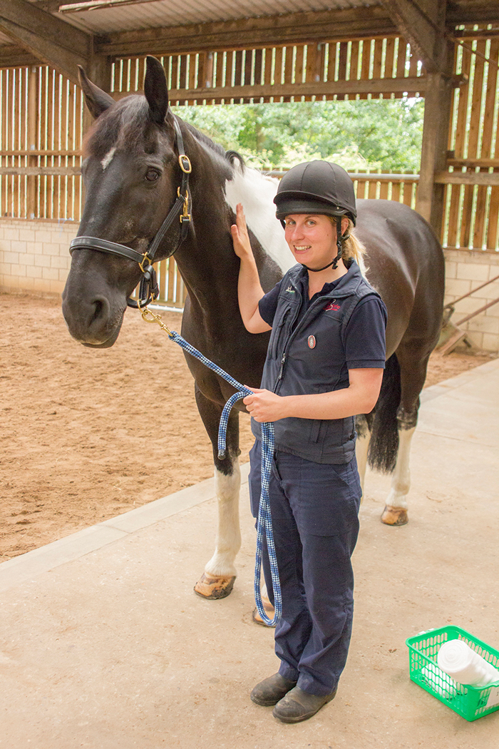
Patient restraint
The RVN could have a role in restraint of the patient for all of the procedures mentioned. It is essential that the patient is calm and relaxed at all times, even if sedation is to be administered.
The RVN should ensure adequate methods of restraint are available – for example, head collar, bridle or nose twitch – and used with skill and caution. The RVN should also wear correct PPE – for example, a hard hat and steel toe-capped boots (Figure 1).
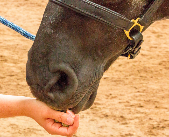
The patient should be kept calm by the RVN talking calmly, and gently stroking the horse to provide reassurance. Food such as treats can be offered as a reward for positive behaviour (Figure 2). It is far better to make the experience a calm and positive one for the horse, as this will make further procedures safer and easier for all involved.
Conclusion
RVNs are not commonly involved in the diagnosis of dermatological conditions in the horse; however, they could clearly take on a number of important roles.
Many samples can be taken by RVNs, such as skin scrapings, acetate tape impressions and hair plucks. Microscopic examination of the samples can also be carried out by RVNs to identify the causal agent. After consultation with the case vet, RVNs can administer treatment and monitor the patient for progress.
The role of owner education should not be overlooked. RVNs can advise owners on the use of medication and methods of infection control. This not only allows the RVN to use his or her expertise, but also helps raise public awareness of the role of the veterinary nurse in practice. RVNs can also assist the case vet with diagnostic procedures, including correctly and safely restraining the patient, as well as clipping and preparing the designated site. RVNs can also provide the correct equipment and apply infection control protocols.
Leave a Reply