Presented are selected cases from the ruminant diagnostic caseload of Axiom Veterinary Laboratories.
Axiom provides a farm animal diagnostics service to more than 300 farm and mixed practices across the UK, and receives both clinical and pathological specimens as part of its caseload. The company is grateful to clients for the cases presented in this article.
The focus for this article is gastrointestinal and nutritional disease in winter 2019-20.
Intestinal parasites
Additionally to numerous diagnoses in young calves, cryptosporidiosis was diagnosed in neonatal orphan lambs.
Infection typically occurs between 4 and 10 days of age, and is transmitted by the faeco-oral route. The oocysts are resistant and widespread environmental contamination can occur – from which lambs acquire infection.
Coccidiosis was confirmed in calves with the identification of pathogenic Eimeria species (Eimeria bovis, Eimeria zuernii and Eimeria alabamensis). Reported signs included illthrift, scour, tenesmus, recumbency and mild pyrexia.
It was also suspected in several other calves, with clinical histories suggestive of coccidiosis where high counts of unspeciated coccidial oocysts were seen in faeces.
Coccidiosis was confirmed in one of five young emaciated Boer goats; a count of 11,100opg Eimeria ninakohlyakimovae being detected in faeces. This species is considered the most pathogenic Eimeria species in goats and can cause widespread denudation of the mucosa in the upper large intestine.
Other pathogenic species seen in high coccidial oocyst counts reported in goat faeces included Eimeria hirci, Eimeria arloingi and Eimeria christenseni.
Gastroenteritis
Parasitic gastroenteritis was detected in several herds – primarily in calves up to five months of age – but the highest count was seen in a weak, recumbent 13-month-old heifer with a faecal strongyle egg count of 36,600epg and an unspeciated coccidial oocyst count of 5,400opg.
High pepsinogen levels consistent with ostertagiosis were also detected in youngstock up to two years of age; signs included illthrift and scour.
Multiple cases of parasitic gastroenteritis were also reported in sheep and goats, with occasional counts exceeding 50,000 strongyle epg. Mortality was reported in some flocks.
In one group of six-month-old lambs, a strongyle egg count of 1,400epg was detected in faeces 10 days post-treatment with a closantel-mebendazole combination, suggesting treatment failure.
Counts of up to 900epg Nematodirus battus were seen in some flocks alongside high strongyle egg counts.
In some sheep and goats, low vitamin B12 levels consistent with concurrent cobalt deficiency were seen alongside high strongyle egg counts. In one flock of seven-month-old to eight-month-old lambs with coughing and scour, Dictyocaulus filaria were also detected – using the Baermann technique – in addition to a high strongyle egg count.
Fasciolosis
Fasciolosis was detected by coproantigen ELISA and microscopy on a number of herds and flocks. In clinical cases, signs reported included malaise, illthrift, milk drop and submandibular swelling.
In one case of suspected treatment failure, an illthrifty Soay adult sheep with bottle jaw had been treated with albendazole two weeks earlier; the flock was on marshy ground and a number of deaths had occurred.
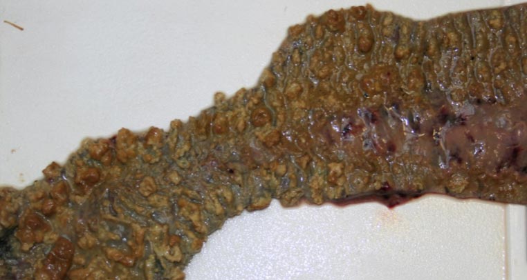
Johne’s disease
Several cases of Johne’s disease were seen in 18-month-old cattle this quarter.
In an interesting case in an older animal, a scouring four-year-old bull in a dairy herd was found to be Johne’s PCR positive and seronegative. Possible explanations included anergy – when an animal overwhelmed by infection is unable to mount an effective immune response – and a false negative serology result (with a sensitivity of 90% for clinical cases; up to 1 in 10 clinically affected animals may give this).
A false positive PCR result – for example, with passage of Mycobacterium avium subspecies paratuberculosis (MAP) through the animal after ingesting an environmental source – may have been less likely due to the reported clinical signs.
In a second case, an illthrifty scouring Friesian cow was seropositive and had a low threshold cycle value for MAP consistent with a high antigenic load in faeces, despite being a low-risk animal on milk serology.
In a third case, a scouring Holstein cow was negative on quarterly milk serology, but highly seropositive on serum ELISA. Milk serology is not as sensitive as blood testing; sensitivity was 72% compared to blood in one study using the same ELISA.
Goats and sheep
Johne’s disease was confirmed in a number of goats on serology and PCR.
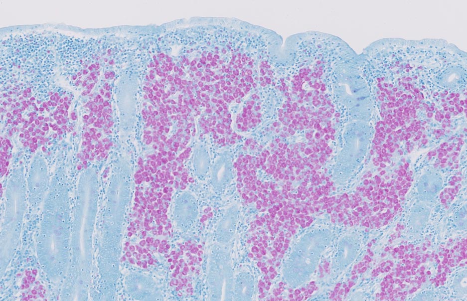
It was also diagnosed on postmortem examination (PME) in a 12-year-old pygmy goat with weight loss, malaise, anaemia and some scour. Histopathology revealed granulomatous inflammation in the small and large intestine, draining lymph node and liver. Lesions were typical of those seen with Johne’s disease due to MAP – and on a Ziehl-Neelsen stain, numerous clusters of short acid-fast bacilli were present within mucosal macrophages, with smaller numbers present within the submucosa of the small intestine.
Dissemination of infection from the intestine to draining lymph nodes – and, subsequently, via the portal system to the liver – is commonly seen in well-established cases.
Johne’s disease also was confirmed in a three-year-old Charollais ram that had a high strongyle count (1,800epg) – and in one flock screen, two of three pooled faecal samples were positive for Johne’s on PCR.
Salmonellosis
Salmonella enterica serovar Dublin was isolated on 22 occasions in the past quarter from the faeces of calves and cows. In most cases, a history of scour existed, which was occasionally haemorrhagic or contained intestinal casts.
In one group of two-week-old dairy calves, ear tip necrosis and meningitis also had been reported, with 3 of the previous 10 calves dying.
S enterica serovar Typhimurium – and variants of this serovar (Copenhagen and 4, 12:i:-) – were isolated on seven occasions from cows, beef finishers and calves; affected animals scouring where reported. In one 17-month-old male Charolais beef finisher, severe diphtheritic enteritis was found at PME.
S enterica serovar Mbandaka was isolated on seven occasions, again from calves and cows, described as haemorrhagic in six cases.
S enterica serovar Berta was isolated from a nine-month-old chronically scouring calf, which also had a strongyle egg count of 1,000epg. This serovar is rarely reported in cattle, is more typically isolated from poultry, and has been associated with outbreaks of illness and diarrhoea in people.
Another rare serotype in cattle – S enterica serovar Uganda – was isolated from a three-year-old dehydrated, pyrexic dairy heifer with watery scour containing gut lining that had calved six days previously. Outbreaks in humans have been associated with papaya and pork products. Further cases were subsequently confirmed in the herd.
Three other less frequently encountered serovars were also reported:
- Salmonella 6,7:-:enz15 was isolated from a scouring cow.
- Salmonella 3,10:e,h:- was isolated from the faeces of scouring Holstein calves.
- Salmonella 6,7:r:- was isolated from the faeces of a five-year-old Holstein-Friesian cow that was scouring post-calving.
Salmonella 61:-:1,5,7 – a variant of S enterica subspecies diarizonae – was isolated from a duodenal swab taken from a Texel ram that was lethargic and inappetant prior to death, and, on PME, had enlarged mesenteric lymph nodes and odoriferous ruminal contents, but no visible scour.
This was the fourth of 10 rams to succumb in this manner and some of those affected were in poor body condition.
Abomasitis
Sarcina-associated abomasitis was diagnosed on histopathology in a nine-day-old Jersey calf that had a history of intermittent bloating before being found dead.
Gross PME found evidence of an emphysematous abomasitis. Sarcina-like bacteria were seen on histopathology, together with large bacilli, most likely clostridia.
The condition is thought to be a mixed infection of Sarcina and clostridial species, including Clostridium perfringens and Clostridium fallax. Predisposing factors include poor hygiene, recent and rapid changes in feeding practices, and feeding milk at different temperatures.
Bacterial abomasitis
Acute bacterial abomasitis was confirmed on histopathology in an 18-month-old ewe that died after a period of lethargy and scour.
On PME, the abomasal mucosa was haemorrhagic and morphology of bacteria seen on a Gram stain most closely resembled Trueperella pyogenes or Listeria species.
T pyogenes is commonly found on mucosal surfaces and can act as a secondary pathogen, while Listeria monocytogenes has been implicated in cases of abomasitis – usually with concurrent severe necrotising typhlitis – and is normally associated with feeding of silage with a high listerial count.
Mucosal disease
Bovine viral diarrhoea viraemia was detected by antigen ELISA in a 13-month-old Limousin-cross bull beef animal with a history of watery diarrhoea and tenesmus for four days; this was, therefore, likely to be a case of mucosal disease.
A second case was suspected in a thin 14-month-old viraemic beef finisher with scant watery, bloody scour and pyrexia (40.5°C); and a third case in an off-colour, scouring three-month-old viraemic Limousin calf.
Enterotoxaemia
Type D clostridial enterotoxaemia was suspected with the detection of epsilon toxin in intestinal contents of a dead female goat – 1 of approximately 20 in each of two groups of 100 that had profuse watery scour, with four or five animals dying acutely.
The goats were being pushed hard for yield, but had received Lambivac.
A second case was detected in a four-year-old golden Guernsey goat with sudden onset watery scour containing mucosa.
Vitamins and minerals
Magnesium
Following a cow – three months in calf – being found dead with evidence of paddling, sampling of other cows detected hypomagnesaemia in 4 out of 10, while several others had marginal magnesium levels.
Cobalt
Cobalt deficiency was detected with demonstration of low vitamin B12 levels in several sheep flocks and goat herds; signs where reported included illthrift, scabbing around eyes and ears, scour, and weakness. Some cases were seen together with selenium deficiency.
Copper
Low copper levels were reported in suckler and dairy herds, and in sheep flocks – some cases together with low vitamin B12 or glutathione peroxidase (GSH-Px) levels. Where clinical signs were reported, these included illthrift and, in cattle, poor fertility.
Copper deficiency also was confirmed on liver copper analysis in a dairy cull cow (114µmol/kg dry matter [DM]; reference interval [RI] 314 to 7,850).
A copper level of 31.3µmol/l (RI 9 to 19) was suspected to be consistent with copper poisoning in a sick six-year-old ram with brown mucous membranes and dark mucoid faeces.
Selenium
Selenium deficiency alone was diagnosed in several herds and flocks, primarily through detection of low GSH-Px levels. Clinical signs, where reported, included illthrift, weakness and lethargy.
One 18-month-old Charolais beef finisher, that had been housed for four days, became ataxic on the hindlegs, progressing to recumbency. A low liver selenium level of 0.31mg/kg DM was found (RI 0.9 to 1.65) and it was likely the clinical signs were due to white muscle disease.
The animal also had a low liver copper level (112µmol/kg DM).
Manganese
Manganese deficiency was again reported on a dairy farm with known low manganese levels in the soil. Attempts had been made to supplement the herd’s ration, but this had not increased manganese levels in the stock.
No evidence of chondrodystrophy was present in calves, but herd fertility was suboptimal; manganese deficiency may have been a contributory factor, as depressed or delayed oestrus – and poor conception rates – have been associated with experimental manganese deprivation in cattle, sheep and goats.
Low levels also were found in a screen of a herd with a five-year history of manganese deficiency, where clinical cases of chondrodystrophy had been seen. It had been supplemented with manganese for the past six weeks.
A number of other herd screens had low manganese levels, and an individual case of manganese deficiency was diagnosed in a seven-month-old calf with a stiff gait and slightly swollen distal limb joints.
Vitamins
In a herd with a higher-than-acceptable incidence of weak calves and navel infections, four out of six calves sampled had low vitamin E levels.
Colostrum is the main source of vitamin E for neonatal calves, so this may have been a sign of inadequate colostral absorption. It was advised zinc sulphate turbidity or total protein levels were checked in calves aged two to seven days to assess colostral intakes.
Low vitamin A levels were found in all four samples taken from weaned Holstein-Friesian calves that had become recumbent for no clear reason.
Vitamin A and E, and selenium deficiency were diagnosed in 14-month-old Limousin fattening cattle that were going off their legs.
Low vitamin D levels were detected in two of four four-month-old Holstein calves from a herd with a high incidence of fractured legs in youngstock. Vitamin D deficiency had been confirmed last winter and the animals were now a higher rate of vitamin D supplementation.
Other cases
Low T4 levels were seen in five out of six samples on a cull ewe screen, suggestive of potential iodine deficiency.
Suboptimal urea levels were detected in all seven blood samples taken from a group of thin sucklers, consistent with a lack of rumen-degradable protein caused by either inadequate levels in the diet or insufficient intake.
Suboptimal urea levels also were detected in:
- all five dairy-bred heifers with illthrift and poor fertility in a second herd
- in three Sussex heifers/cows in a third herd on barley, silage and a protein mix, and suffering from illthrift post-calving
Leave a Reply