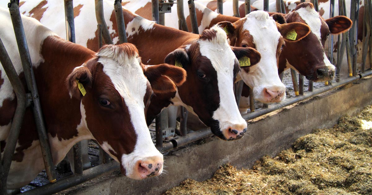
Presented are selected cases from the ruminant diagnostic caseload of Axiom Veterinary Laboratories.
Axiom provides a farm animal diagnostics service to more than 300 farm and mixed practices across the UK, and receives both clinical and pathological specimens as part of its caseload. The company is grateful to clients for the cases presented in this article.
The focus for this article is respiratory disease in spring 2019.
Infectious bovine rhinotracheitis (IBR) was detected by PCR in a conjunctival swab taken from one of 40-plus affected calves out of a group of 100, all younger than seven days – with conjunctivitis, swollen eyelids, purulent to watery ocular discharge, and occasional pyrexia (39.5°C to 40°C).
IBR also was detected by PCR in:
- a trachea sample from a two-week-old Holstein-Friesian calf
- a lung from a calf younger than one month (also with fibrinosuppurative pleurisy)
- three post-weaned calves with pyrexia, oculonasal discharge and increased upper respiratory tract sounds
- beef finishing cattle in two different herds
- a six-year-old cow with acute respiratory distress in a herd where an outbreak of ocular and nasal discharge occurred three weeks after buying in cattle
It was also confirmed with rising titres on paired IBR gE serology in an adult dairy cow, and in four indoor beef finishers with acute respiratory infection in different herds.
Respiratory syncytial virus
Several cases of respiratory syncytial virus (RSV) also were diagnosed on PCR. The virus was detected in lung samples from:
- a three-month-old calf
- an eight-month-old Friesian calf that also had evidence of an acute fibrinosuppurative and necrotising bronchopneumonia on histopathology, indicating a bacterial component to the pneumonia
- a three-month-old calf that died a day after developing severe respiratory signs
It was also detected by PCR on pooled nasal swabs from four-week-old calves with nasal discharge and increased respiratory rates, and in an adult suckler with extensive SC emphysema.
Rising titres to RSV were detected on paired serology in a preweaned dairy calf and in suckler calves in two herds, and rising titres to both RSV and IBR were seen in one each of three cows in a herd with respiratory signs and pyrexia.
Pasteurellosis
Pasteurellosis was diagnosed on culturing of Mannheimia haemolytica or Bibersteinia trehalosi and/or histopathology in lung samples received from lambs in several flocks.
Although many animals were found dead, one clinical case was a six-week-old lamb with severe depression, and increased respiratory sounds and effort; and a second was a four-week-old lamb that died after a period of illthrift in a flock where seven other lambs had died acutely. Gross signs included fibrinosuppurative pneumonia, pleurisy and pericarditis.
In one case, histopathology suggestive of either M haemolytica or B trehalosi septicaemia was found in a reportedly vaccinated, thin ewe lamb, with an acute embolising bacterial pneumonia and multifocal bacterial emboli in the kidneys.
In other species, pasteurellosis was suspected on histopathology in a group of 10-day-old goat kids that died five days after arrival on farm. It was also confirmed on culture of either Pasteurella multocida or M haemolytica from lung and/or lung histopathology.
It was also diagnosed in one of two dead beef finishing bulls in which 8 of the 20 in the group were looking poor after being recently moved, and in a 10-day-old calf that died after acute onset of respiratory signs, with cranioventral lung consolidation seen on postmortem examination.
Mycoplasma bovis
Mycoplasma bovis was detected by PCR in the lung of a preweaned Limousin-cross calf, one of a number to have died from pneumonia. On postmortem examination, the lungs contained multifocal small abscesses.
In a second herd, in which 50% of calves were dying with scour; focal lung abscessation was seen on examination of a 12-week-old calf. Histopathology revealed severe chronic active pneumonia plus caseous abscessation consistent with infection with M bovis and other bacteria.
A similar histopathological pattern was seen in the lung of a three-month-old suckler calf, while active seroconversion to M bovis was detected in two cases of pneumonia – a post-weaned dairy calf and a dairy cow – in different herds.
Atypical pneumonia
Atypical pneumonia was diagnosed in several flocks. This disease has a multifactorial aetiology.
In some cases, the presence of bronchiolitis obliterans suggested an initial viral insult – such as parainfluenza-3 virus or RSV – and, in many cases, histopathology suggested mycoplasmal involvement (typically Mycoplasma ovipneumoniae).
Some cases also had a significant suppurative component on histopathology consistent with an additional bacterial aetiology – for example, M haemolytica, B trehalosi, P multocida, staphylococci or streptococci.
The condition is more frequently seen in post-weaned lambs, which present with a soft cough and illthrift, but cases in much younger lambs appear to be increasing.
Lungworm
Lungworm was confirmed – via the Baermann technique – in yearling heifers and calves in different herds.
The sensitivity of the Baermann test is very much affected by sample quality, as lungworm larvae may die in transit to the laboratory, particularly in warm ambient temperatures.
Faeces samples should be immediately refrigerated then sent to the laboratory the same day they are passed by the animal.
Ovine pulmonary adenocarcinoma
Ovine pulmonary adenocarcinoma was diagnosed on histopathology on five occasions.
In one case, it caused one of six deaths in a flock of 120 Welsh Mountain ewes where older animals were primarily affected, and exhibiting neurological signs and bottle jaw.
Concurrent copper and selenium deficiency were detected on liver biochemistry, and a worm egg count of 1,600epg was detected on an in-house faecal egg count.
In two other flocks, evidence of secondary bacterial pneumonia was also present.
Seroconversion to maedi-visna was detected in several flocks, including a flock in which ewes were not letting down any milk. Two ewes also had lung histopathology suspicious of maedi-visna.
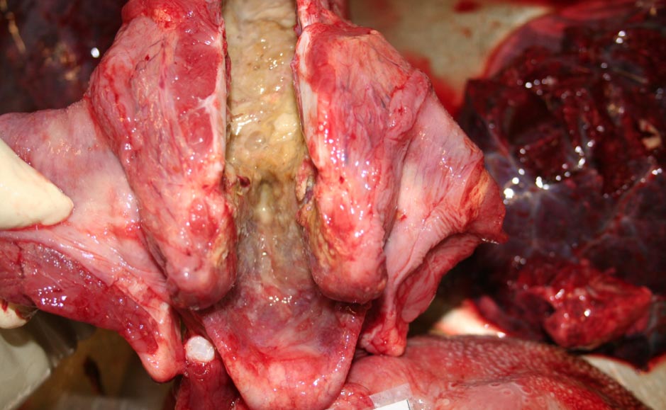
Laryngeal chondritis
Laryngeal chondritis was confirmed in a yearling Texel tup that died suddenly.
Focal ulceration of the laryngeal mucosa occurs with secondary bacterial infection and involvement of deeper structures, including skeletal muscle and laryngeal cartilage.
Animals usually present with stertor and will commonly develop acute respiratory signs often associated with laryngeal oedema and occlusion, leading to death.
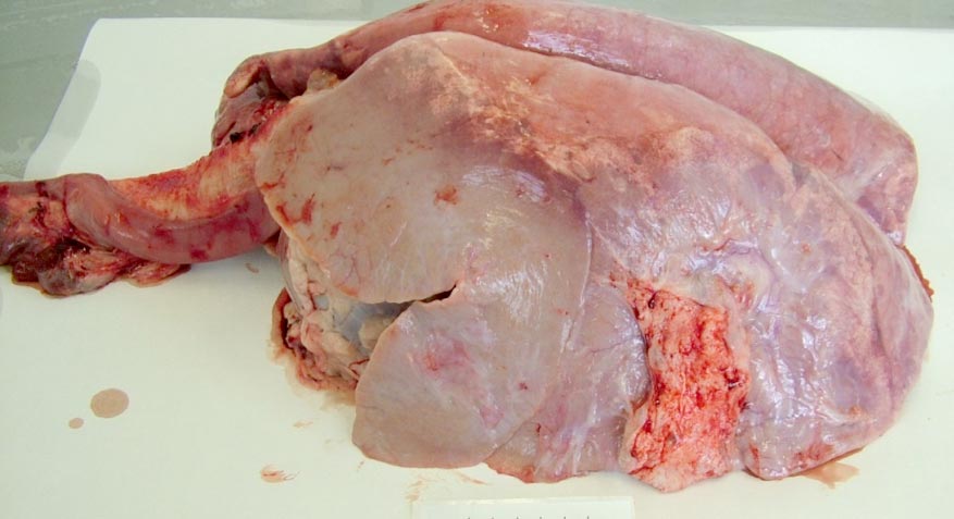
Bronchopneumonia
Chronic bronchopneumonia was detected on histopathology of the lung of a four-year-old East Friesian ram with a history of coughing and extensive pleurisy on postmortem examination.
Renal amyloidosis was also present, which usually occurs as a sequel to chronic inflammatory diseases – such as pneumonia – and to neoplastic disease.
Pulmonary thromboembolism
Pulmonary thromboembolism with suppurative pneumonia was the cause of sudden onset of respiratory signs and pyrexia in a Holstein-Friesian cow.
This is most commonly associated with liver abscesses adjacent to the caudal vena cava, resulting in thrombosis. Other potential sites include right atrioventricular valve endocarditis, uterine vein phlebitis and milk vein phlebitis.
Leave a Reply