Presented are selected cases from the ruminant diagnostic caseload of Axiom Veterinary Laboratories.
Axiom provides a farm animal diagnostics service to more than 300 farm and mixed practices across the UK, and receives both clinical and pathological specimens as part of its caseload. The company is grateful to clients for the cases presented in this article.
The focus for this article is systemic and miscellaneous diseases in late summer/autumn 2019.
Among the many cases of bovine viral diarrhoea (BVD) viraemia was an eight-week-old Holstein-Friesian calf found to be BVD viraemic at birth on tag and testing, which was again positive for BVD antigen ELISA on a serum sample – confirming persistently infected (PI) status.
Surprisingly, the calf also had a strongly positive titre on BVD serology. PCR testing at APHA Penrith confirmed it was BVD type one and it was suspected the antibody titre was due to unusually high levels of maternal antibody.
In a second case, a recumbent three-week-old Hereford bull with navel and joint ill had a PCR cycle threshold (Ct) value of 25, likely to be consistent with PI status.
In a third case, BVD viraemia with a PCR Ct value of 26 – again suspicious of PI status – was confirmed in an 18-hour-old Hereford-cross calf that was blind, unable to stand and shaking, strongly suggestive of cerebellar hypoplasia.
Cases of mucosal disease included:
- a Simmental steer with condition loss and scour, with blood and sloughed mucosa found on rectal examination
- a four-year old, BVD-vaccinated Limousin stock bull that was tucked up, scouring and had a thickened rectal wall on palpation
- a recently purchased Limousin-cross beef finisher with wasting, scour and nasal crusting
Border disease and malignant catarrhal fever
Border disease viraemia was confirmed by PCR in a number of sheep flocks.
In one submission, seven border disease-viraemic animals were found in a large Swaledale flock screen of 555 animals using a combination of pooled PCR, individual PCR and serology.
Malignant catarrhal fever was confirmed in several animals – primarily by PCR, but occasionally on serology. Signs included:
- oral and nasal ulceration
- nasal oedema
- mucopurulent nasal discharge
- conjunctivitis
- corneal oedema
- ocular discharge
- vaginal ulceration
- weight loss
- scour
- pyrexia (higher than 41°C)
- coughing
Pulpy kidney and louping ill
Pulpy kidney was the likely cause of sudden death in a four-month-old Jacob lamb, with Clostridium perfringens alpha and epsilon toxins detected on ELISA in small intestinal contents.
It was also suspected when epsilon toxin was detected by ELISA in small intestinal contents from one of three fattening goats that died acutely, and from ileal contents in one of four easy-care lambs to die over a four-day period; a fibrinous pericardial effusion was present in both animals.
Louping ill was confirmed on haemagglutination inhibition test serology in two 18-month-old hardy speckled face gimmers showing neurological signs (ataxia and slight tremor). Ticks could not be seen on the sheep, but a history of louping ill in hill ewes existed in the area.
Babesiosis and tick-borne fever
Several cases of babesiosis were diagnosed in cattle on haemoparasite screens, with characteristic inclusions in erythrocytes.
In one case, a history of redwater had been present in a group of more than 15 cows in a valley; they had been brought inside more than 160 days after housing. One animal developed pyrexia (40°C) and haemoglobinuria, and babesiosis was confirmed on a blood smear. It was suspected ticks may have survived in the bedding.
In a second case, babesiosis was confirmed in the third cow to become sick and recumbent in a batch of sucklers bought two months earlier – one cow dying a week after arrival and the second a few weeks later. No scour or jaundice were seen, but the cow had marked azotaemia, presumably due to haemoglobinuric nephrosis secondary to haemolysis.
Tick-borne fever was confirmed when characteristic inclusions were seen in leukocytes in a cow with pyrexia (42.2°C), excess salivation, photophobia, some epiphora and mild nasal discharge. Other cows had been affected over the previous 7 to 10 days and had recovered.
Salmonellosis
Systemic salmonellosis was reported on a number of occasions.
Salmonella typhimurium was isolated from a faecal sample from a 15-month-old bull with purulent polyarthritis after fighting with another bull, the animal going on to scour with blood clots and become recumbent.
Salmonella 4, 5, 12:i:- was isolated from a 10-month-old dehydrated, bloated barley beef finisher in a herd with a history of bloat and pneumonia.
Salmonella enterica subspecies enterica serovar Dublin was isolated from a swab taken at the postmortem examination (PME) of a two-month-old Holstein calf with hepatomegaly, jaundice and signs of septicaemia; and from intestinal contents from a recumbent, pyrexic 14-day-old Holstein-Friesian calf that was euthanised and found to have pneumonia on PME. It was also isolated from turbid joint fluid taken from one of a group of poorly doing three-month-old Holstein-Friesian calves.
Tumours
Adenocarcinoma was diagnosed as the cause of visceral plaques and ascites in a seven-year-old ewe.
This is a common tumour of older sheep that usually arises from the intestine. Transcoelomic spread results in multifocal firm, white, slightly raised plaques adherent to visceral surfaces, and occlusion of diaphragmatic lymphatics by neoplastic emboli may result in marked ascites.
Although a specific aetiology has not been established, cases can often be seen in areas with extensive bracken, suggesting a possible link.
A poorly differentiated sarcoma was confirmed on histopathology of a discharging mass on the thorax of a two-year-old male pygmy goat.
A vaginal mass removed from a Galloway cow was determined to be a squamous cell carcinoma on histopathology. These tumours often occur on areas of non-pigmented skin that can be exposed to ultraviolet light, resulting in neoplastic transformation.
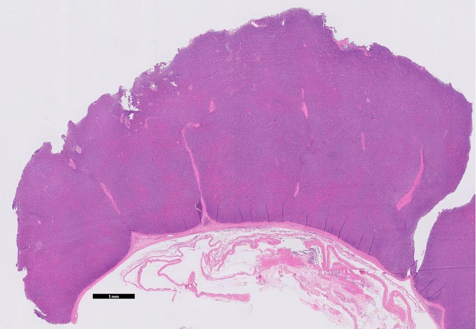
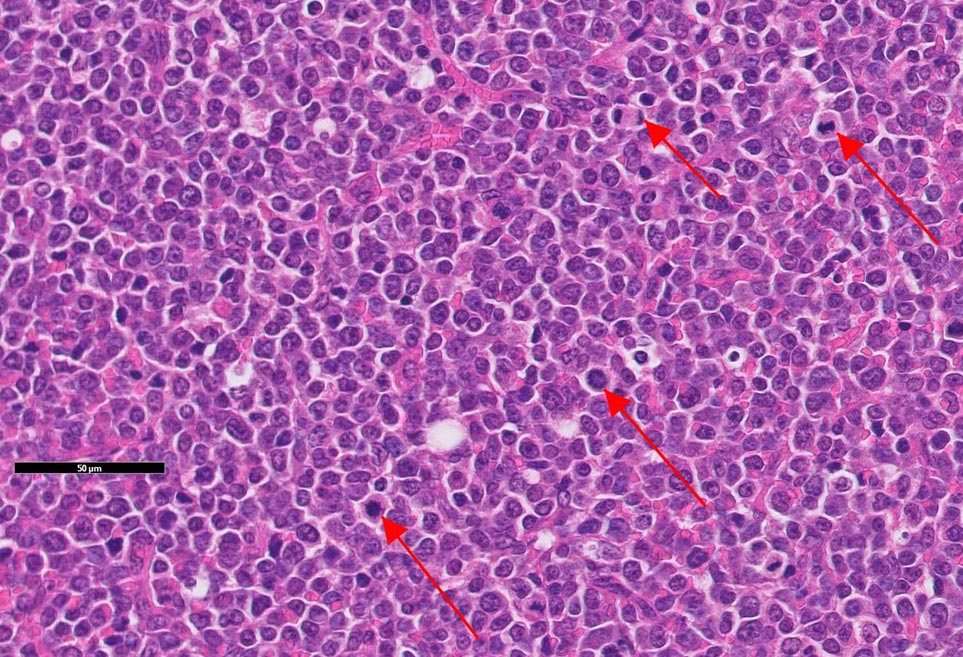
Lymphoma was diagnosed on histopathology of a 40cm diameter mass attached to the pericardium of an 18-month-old Aberdeen Angus bull that had developed acute respiratory signs a few hours before dying. Additional findings on PME included extensive inflammation of the upper respiratory tract, haemothorax and enlarged thoracic lymph nodes.
Lymphoma also was the cause of a nodular cervical mass in one calf, and generalised lymph node enlargement and pyrexia in another calf in different herds. The second case was consistent with the juvenile multicentric form of sporadic bovine lymphoma; this most commonly occurs in calves between three months and months of age, but may be present at birth.
As none of the animals were older than two years of age, the finding did not need to be reported to the APHA to investigate the possibility that enzootic bovine leukosis virus was the cause of the neoplasia.
Fat necrosis
PME of a 15-month-old Holstein-Friesian bullock revealed haemorrhagic loops of colon bound up in a fatty abdominal mass. Histopathology of mesenteric fat was suggestive of massive fat necrosis.
In cattle, this is characterised by multifocal masses of fat necrosis of different sizes within the abdominal cavity that can entrap the intestine, with subsequent compression and obstruction.
It is normally seen in older animals, particularly Channel Island breeds, but the pathogenesis remains unclear; it may be associated with alterations in lipid metabolism and dietary factors, including ingestion of feeds with high levels of long-chain saturated fatty acids.
Elevated enzymes
A number of cases of suspected hepatogenous photosensitisation were reported in suckler herds, affected animals having raised liver enzymes.
Clinical signs reported included:
- pyrexia
- serous and purulent ocular discharge
- aural discharge
- nasal discharge
- ulcerated and peeling muzzles
- pyoderma on ears
- sloughing mucocutaneous junctions
- ulcerated, crusting udders and teats
Screening of a group of apparently healthy, post-weaned Holstein-Friesian calves revealed high glutamate dehydrogenase (GLDH) levels (36U/L to 438U/L; reference interval less than 25), with some calves also having neutrophilia and/or monocytosis suggestive of infection/inflammation.
We have seen a number of similar cases in recent months in young calves that were reported to be clinically healthy. It is possible this pattern could occur with (subclinical) ruminal acidosis.
A similar high GLDH pattern has been reported in preweaned calves that had acidosis due to milk indigestion.
Caprine arthritis encephalitis and caseous lymphadenitis
Screening of four male goats in a herd found two were seropositive for caprine arthritis encephalitis – and, therefore, potential shedders of the virus – and three were seropositive for caseous lymphadenitis (CLA).
Although the CLA ELISA assay is not yet fully validated for use in goats, it is believed it should perform as well as it does in sheep, in which it has 98% specificity. However, vaccinated animals can test positive in the CLA ELISA.
CLA also was confirmed on serology in three golden Guernsey goats with parotid and mandibular abscesses, and on culture of abscesses in two rams from different flocks.
Orf
Orf was identified by histopathology as the cause of ulcerated lesions on the lips and dental pad of an ill-thrifty, six-month-old Border Leicester ewe lamb. It also had severe chronic bacterial bronchopneumonia.
The chronic nature of the orf lesions was probably due to poor cell-mediated immunity in a debilitated animal resulting in viral persistence, allowing more proliferative lesions to be produced.
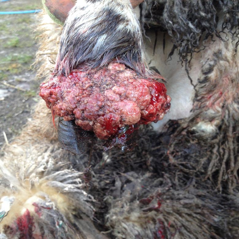
A second case of orf was diagnosed by PCR in a five-month-old, ill-thrifty lamb with lesions around the muzzle, and a third case in a lamb with two large, weeping lesions on the front and hind right feet attached at the coronary band. The description suggested the latter may have been a case of strawberry foot rot, with Dermatophilus congolensis as a secondary pathogen.
Sheep scab was confirmed on microscopy of wool plucks and skin scrapes in one flock.
Miscellaneous
Cardiac blackleg (clostridial myositis) was diagnosed on histopathology in a well-conditioned, six-month-old Hereford-cross calf that died acutely with no premonitory clinical signs.
On PME, the heart was found to be enlarged and haemorrhagic. Youngstock were not vaccinated against clostridial disease.
Corynebacterium renale, a known cause of pyelonephritis and cystitis in cattle, was isolated from the urine of a cow that was off-colour and had milk drop over a five-day period.
Trueperella pyogenes was isolated from submandibular abscesses in a Texel-cross ewe and in a one-year-old ram in different flocks. It was also isolated from swabs taken from injection site reactions (abscessation/cellulitis) in two Texel-cross lambs after administration of a multivalent clostridial/Pasteurella vaccine.
Peptoniphilus (formerly Peptostreptococcus) indolicus was isolated on anaerobic culture of the same swabs. Members of this genus have been involved in the aetiology of summer mastitis in cattle and associated with soft tissue infections in humans. Therefore, its isolation may have been of significance.
Actinomyces hyovaginalis was isolated from a submandibular abscess in an 18-month-old Texel tup. This isolate is an opportunist pathogen most typically isolated from pigs, but at least one report exists in the literature of its isolation from sheep.
Profuse growths of Staphylococcus aureus were isolated from swabs taken from acute onset crusting, purulent lesions on the nares of a four-month-old calf, and a two-year-old beast in a suckler herd. S aureus infections usually occur secondary to trauma – for example, at trough feeding.
Treponema phagadensis was detected by PCR on a deep swab taken from a swelling on the mid cannon bone of one leg of an acutely lame Holstein-Friesian; 12 of 240 cattle were similarly affected. This treponeme is associated with digital dermatitis in cattle.
High titres to Erysipelothrix rhusiopathiae were detected in four or five four-month-old lambs with joint ill, consistent with recent exposure, suggesting this was the cause of the joint ill.
Mycoplasma conjunctivae was detected by PCR in a group of newly purchased ewe lambs with pink eye.
Leave a Reply