Insect bite hypersensitivity (IBH) is a chronic, recurrent and seasonal (spring, summer and autumn) dermatitis that is known to affect 5% to 50% of horses, dependent on their location, breed and genetics (Schaffartzik et al, 2012).
The pathological state is an allergic, pruritic dermatitis predominantly caused by IgE-mediated type-one hypersensitivity reaction to the antigens present within the saliva of Culicoides species.
Atopic dermatitis (AD) is another common dermatological condition that is again associated with IgE-mediated hypersensitivity to various allergens.
Clinical presentation
In IBH cases, horses are most affected in the summer, although disease can also be seen in both spring and autumn.
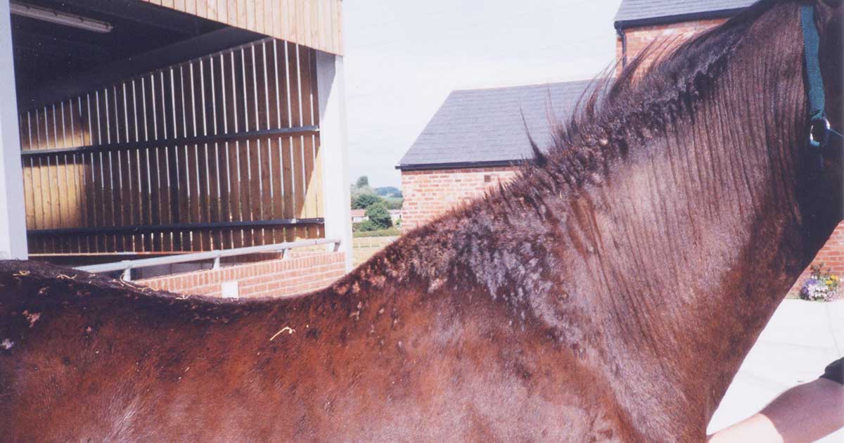
Horses will show severe pruritus of the mane (Figure 1) and tail area (Figure 2), while, to a lesser degree, some will be affected along the ventral midline and face. Often, clinical signs will start with papules, urticaria and tufted hair or changes in hair structure (Figure 3).
These changes can then progress to self-excoriation due to the severity of the pruritus. Winter normally leads to relief as the Culicoides are no longer present in the environment.
Patients with AD can be affected year-round, depending on the allergen involved, but frequently horses will appear worse in periods of high-allergen load (that is, when plants are flowering, for example).
Lesions can appear similar to IBH, but can affect any part of the body – depending on the allergen involved – and the horses will frequently be pruritic.
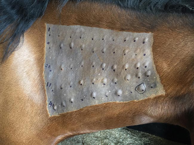
Diagnosis of IBH is frequently made on the clinical signs alone, as they can be pathognomonic for the disease. Intradermal (Figure 4) and serological tests have been trialled, but the results have been variable – with cases of both false-positive and false-negative results occurring due to the use of whole Culicoides for the antigen, rather than the saliva proteins themselves (Quinn et al, 1983; Sloet van Oldruitenborgh-Oosterbaan et al, 2009).
AD conversely requires the use of intradermal skin tests to guide the diagnosis, as well as assessment of the environment and the allergens the horse is exposed to (Rendle et al, 2010). Biopsy, in both cases, can be informative, as it will ensure no other aetiologies are leading to the clinical signs. Also, it can confirm a high number of eosinophils, likely indicative of an allergic response.
If biopsies are taken, it is also wise to culture the samples to ensure no secondary infection is present within the skin. Particular attention should be paid to growth of Staphylococcus aureus for evidence of meticillin resistance.
Treatment
For both syndromes, treatment and prevention will be lifelong, and will require intense management on the part of the owner. A multimodal approach is the most likely to secure comparative success in either case.
When considering IBH, treatment should include the use of topical insect repellents or blankets, medications – including topical treatments, steroids and antihistamines – and stabling at dawn and dusk. Immunotherapy should also be considered.
Peeters et al (2014) found stabling alone resolved clinical signs in 59% of horses, while blankets did so in 49% of cases. This confirms that multiple treatment protocols should be used to achieve the best success rate; treatment should not be based on a single method.
When formulating a treatment protocol with the owners, it is helpful to consider a three-tier system to ensure all aspects of management are covered. The three tiers should include:
- control of the pruritic response to prevent self-trauma
- resolution of infections and epithelial trauma secondary to self-trauma
- prevention of further exposure to Culicoides midges
Control of the pruritic response can be done with either systemic or topical medication. Systemic steroids can be used, with oral prednisolone (1mg/kg by mouth every 24 hours) being the treatment of choice initially.
If a limited response is seen, dexamethasone (0.1mg/kg IM every 24 hours) can be trialled. Antihistamines are of limited use in most cases, but can be used as an adjunctive therapy.
Use of topical treatments, including spray hydrocortisone or cream hydrocortisone/triamcinolone, has its place, but will only be efficacious when limited hair is present.
It should be highlighted that steroids can be used palliatively to break the itch-scratch cycle, but they will not be curative while exposure to the midges is still ongoing. Topical treatment can also include antimicrobial shampoo if a secondary infection has occurred following self-excoriation.
Systemic antibiotics might be warranted in the most severe of cases, but a sensible antibiotic stewardship approach should be taken to their use.
The final tier prevention of further exposure is frequently the most challenging, and should include multiple different techniques. These can include:
- assessing and adjusting horses’ turnout
- insect repellents
- stabling at dawn and dusk, when the midges are at their highest prevalence and feeding the most
- rugs specifically designed to reduce exposure to midges
- the use of fans in the stable to reduce the ability of Culicoides to fly in (although ceiling fans have a limited effect on the number of midges present in the stable)
- mosquito netting that can be placed over doors and windows
- reducing the buildup of standing water during the spring period
Turnout must be addressed, with proximity to hedgerows or static water taken into consideration as these increase the frequency of clinical signs due to the increased burden of midges. Changing the habitat is, therefore, essential.
Chemical repellents or insecticides could be effective in reducing the biting rate or even the population size in an environment. Baker et al (2015) found that pyrethroid-based insecticides, which are available in the UK, caused 100% mortality in exposed Culicoides, so the application of these products to rugs may be very beneficial in controlling the population.
Immunotherapy has little effect on cases of IBH, due to the use of whole Culicoides antigen rather that proteins directly from the saliva of the Culicoides.
Various rugs are available on the market that will reduce the exposure of the horse to midges and will dramatically reduce the incidence of disease. They are a pivotal, if frustrating, part of the treatment protocol.
Following diagnosis of AD via an intradermal skin test, it is possible to use immunotherapy to reduce the clinical signs, and, hopefully, resolve the pruritus. Stepnik et al (2012) evaluated 54 cases of AD treated with immunotherapy, and showed 84% of cases had a reduction in clinical signs and 59% required no further treatment beyond the immunotherapy.
Limited reactions to the immunotherapy were seen, although the author has experienced one horse having a severe pruritic episode following administration that required hospitalisation. Owners should, therefore, be aware of this risk.
Alongside immunotherapy, cases will require removal of allergens where appropriate, and intermittent use of steroids or antihistamines, as described for IBH. Sometimes moving from the current stabling/field can reduce the rate of occurrence.
Conclusion
It is clear to see the management and treatment of IBH and AD are, and will continue to be, challenging. This is mostly due to the limited effect of each treatment, the individualised care and advice that is required for each case, as well as the intense nature of care owners must provide. Often, client compliance can be poor due to the intensive nature of the care – and, therefore, ongoing veterinary support will be required for those cases.